Download
1 / 205
2.07k likes | 2.43k Vues
An Introduction to the Lymphatic System and Immunity. Pathogens Microscopic organisms that cause disease: Viruses Bacteria Fungi Parasites Each attacks in a specific way. 22-1 Overview of the Lymphatic System. The Lymphatic System Protects us against disease

E N D
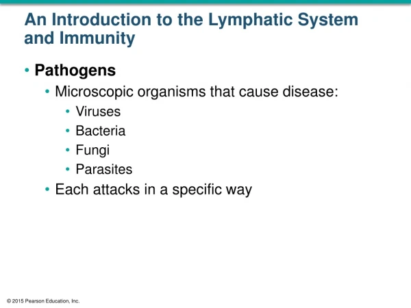
An Introduction to the Lymphatic System and Immunity • Pathogens • Microscopic organisms that cause disease: • Viruses • Bacteria • Fungi • Parasites • Each attacks in a specific way
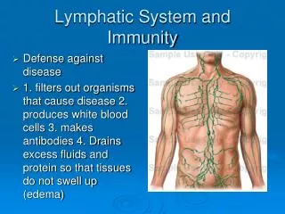
22-1 Overview of the Lymphatic System • The LymphaticSystem • Protects us against disease • Lymphatic system cells respond to: • Environmental pathogens • Toxins • Abnormal body cells, such as cancers
22-1 Overview of the Lymphatic System • Specific Defenses • Lymphocytes • Part of the immuneresponse • Identify, attack, and develop immunity • To a specific pathogen
22-1 Overview of the Lymphatic System • The ImmuneSystem • Immunity • The ability to resist infection and disease • All body cells and tissues involved in production of immunity • Not just lymphatic system
22-1 Overview of the Lymphatic System • Nonspecific Defenses • Block or attack any potential infectious organism • Cannot distinguish one attack from another
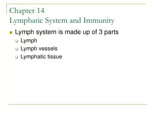
22-2 Structures of Body Defenses • Organization of the Lymphatic System • Lymph • A fluid similar to plasma but does not have plasma proteins • Lymphaticvessels (lymphatics) • Carry lymph from peripheral tissues to the venous system • Lymphoidtissues and lymphoidorgans • Lymphocytes, phagocytes, and other immune system cells
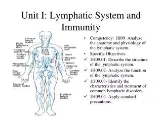
Lymph Lymphocyte Figure 22-1 An Overview of the Lymphatic System (Part 1 of 2). Lymphatic Vesselsand Lymph Nodes Cervical lymph nodes Thoracic duct Right lymphatic duct Lymphoid Tissuesand Organs Axillary lymph nodes Lymphatics ofmammary gland Tonsil Thymus Cisterna chyli Spleen Lymphatics of upper limb Mucosa-associatedlymphoid tissue(MALT) in digestive,respiratory, urinary,and reproductivetracts Lumbar lymph nodes
Lymphatic Vesselsand Lymph Nodes Lymphoid Tissuesand Organs Pelvic lymph nodes Appendix Red bone marrow Figure 22-1 An Overview of the Lymphatic System (Part 2 of 2). Inguinal lymph nodes Lymphatics of lower limb
Arteriole Lymphaticcapillary Smoothmuscle Endothelialcells Figure 22-2a Lymphatic Capillaries. Venule Interstitialfluid Lymphflow Blood capillaries Areolar tissue a The interwoven network formed by blood capillariesand lymphatic capillaries. Arrows indicate themovement of fluid out of blood capillaries and the net flow of interstitial fluid and lymph.
Lymphocyte Incompletebasementmembrane Lymphflow To largerlymphatics Figure 22-2b Lymphatic Capillaries. Areolartissue Interstitial fluid Plasma Interstitialfluid Lymphaticcapillary Bloodcapillary b A sectional view indicating the movement of fluidfrom the plasma, through the tissues as interstitialfluid, and into the lymphatic system as lymph.
Artery Vein Figure 22-3a Lymphatic Vessels and Valves. Lymphaticvessel Vein Artery Lymphaticvessel a Towardvenoussystem Lymphaticvalve From lymphaticcapillaries A diagrammatic view of areolar connective tissue containing small blood vessels and a lymphatic vessel. The cross-sectional view emphasizes theirstructural differences.
Lymphaticvalve Figure 22-3b Lymphatic Vessels and Valves. Lymphaticvessel Lymphatic vessel and valve LM × 63 Like valves in veins, each lymphatic valve consists of a pair of flaps that permit movement of fluid in only one direction. b
Left internal jugular vein Brachiocephalicveins Left jugular trunk Right internal jugular vein Right jugular trunk Thoracic duct Left subclavian trunk Right lymphatic duct Right subclavian trunk Left bronchomediastinaltrunk Right subclavian vein Left subclavianvein Right bronchomediastinaltrunk Superior vena cava (cut) First rib (cut) Figure 22-4 The Relationship between the Lymphatic Ducts and the Venous System. Azygos vein Highestintercostalvein Rib (cut) Thoracicduct Drainage of rightlymphaticduct Drainageof thoracicduct Thoraciclymph nodes Hemiazygosvein Parietalpleura (cut) Diaphragm Cisterna chyli Inferior vena cava (cut) Intestinal trunk Right lumbar trunk Left lumbar trunk a The thoracic duct carrieslymph originating in tissuesinferior to the diaphragmand from the left side of theupper body. The smaller rightlymphatic duct carries lymphfrom the rest of the body. The thoracic duct empties into the left subclavianvein. The right lymphatic duct empties into theright subclavian vein. b
Drainage of rightlymphaticduct Drainageof thoracicduct Figure 22-4a The Relationship between the Lymphatic Ducts and the Venous System. a The thoracic duct carrieslymph originating in tissuesinferior to the diaphragmand from the left side of theupper body. The smaller rightlymphatic duct carries lymphfrom the rest of the body.
22-2 Structures of Body Defenses • Lymphedema • Blockage of lymph drainage from a limb • Causes severe swelling • Interferes with immune system function • Lymphocytes • Make up 20–40 percent of circulating leukocytes • Most are stored, not circulating
22-2 Structures of Body Defenses • Types of Lymphocytes • T cells • Thymus-dependent • B cells • Bone marrow-derived • NK cells • Natural killer cells
22-2 Structures of Body Defenses • T Cells • Make up 80 percent of circulating lymphocytes • Main Types of T Cells • Cytotoxic T (TC) cells • Memory T cells • Helper T (TH) cells • Suppressor T (TS) cells
22-2 Structures of Body Defenses • Cytotoxic T Cells • Attack cells infected by viruses • Produce cell-mediated immunity • Memory T Cells • Formed in response to foreign substance • Remain in body to give “immunity” • Helper T Cells • Stimulate function of T cells and B cells
22-2 Structures of Body Defenses • Suppressor T Cells • Inhibit function of T cells and B cells • Regulatory T Cells • Are helper and suppressor T cells • Control sensitivity of immune response
22-2 Structures of Body Defenses • Other T Cells • Inflammatory T cells • Suppressor/inducer T cells • B Cells • Make up 10–15 percent of circulating lymphocytes • Differentiate (change) into plasmacells • Plasma cells • Produce and secrete antibodies (immunoglobulin proteins)
22-2 Structures of Body Defenses • Antigens • Targets that identify any pathogen or foreign compound • Immunoglobulins (Antibodies) • The binding of a specific antibody to its specific target antigen initiates antibody-mediatedimmunity
22-2 Structures of Body Defenses • Antibody-Mediated Immunity • A chain of events that destroys the target compound or organism • NaturalKiller (NK) Cells • Also called largegranularlymphocytes • Make up 5–10 percent of circulating lymphocytes • Responsible for immunologicalsurveillance • Attack foreign cells, virus-infected cells, and cancer cells
Classes of Lymphocytes subdivided into T Cells Approximately 80% of circulatinglymphocytes are classified as T cells. Figure 22-5 Classes of Lymphocytes (Part 1 of 2). differentiate into HelperT Cells CytotoxicT Cells SuppressorT Cells Memory T Cells Cytotoxic T cellsattack foreign cellsor body cellsinfected by viruses. Helper T cells stimulate theactivation andfunction ofboth T cellsand B cells. Suppressor Tcells inhibitthe activationand functionof both T cells and B cells. Memory T cellsare a subsetof T cells that respond to apreviouslyencounteredantigen.
Classes of Lymphocytes subdivided into B Cells NK Cells NK cellsmake up theremaining5–10% ofcirculatinglymphocytes. B cells make up10–15% ofcirculatinglymphocytes. Figure 22-5 Classes of Lymphocytes (Part 2 of 2). Plasma Cells When stimulated, Bcells can differentiateinto plasma cells,which produce andsecrete antibodies.
22-2 Structures of Body Defenses • Lymphocyte Production • Also called lymphopoiesis, involves: • Bone marrow • Thymus • Peripheral lymphoid tissues • Hemocytoblasts • In bone marrow, divide into two types of lymphoid stem cells
22-2 Structures of Body Defenses • Lymphoid Stem Cells • Group 1 • Remains in bone marrow and develop with help of stromalcells • Produces B cells and natural killer cells • Group 2 • Migrates to thymus • Produces T cells in environment isolated by blood–thymusbarrier
Red Bone Marrow One group of stem cells remains inthe red bone marrow, producingdaughter cells that mature into NKcells and B cells. Multipotenthemopoieticstem cell Figure 22-6 The Origin and Distribution of Lymphocytes (Part 1 of 3). Interleukin-7 Lymphoid stem cells Lymphoid stem cells B cells NK cells
Thymus The second group of stemcells migrates to the thymus,where subsequent divisionsproduce daughter cells thatmature into T cells. Figure 22-6 The Origin and Distribution of Lymphocytes (Part 2 of 3). Thymichormones Lymphoid stem cells Production,selection, anddifferentiatiionof T cells Mature T cells Mature T cells
Peripheral Tissues All three typesof lymphocytes circulatethroughout thebody in thebloodstream,establishingimmunity. Antibody-mediated immunity Immune surveillance Cell-mediated immunity Figure 22-6 The Origin and Distribution of Lymphocytes (Part 3 of 3). One type of mature Tcell, called cytotoxicT cells, plays arole in cell-mediatedimmunity. Thesecells attack anddestroy foreigncells or bodycells infectedby viruses. When stimulated, Bcells can differentiate into plasma cells,which produce andsecrete antibodies.These antibodiesattach to pathogens.This starts achain reactionthat leads tothe destructionof the pathogen. NK cells attackforeign cells, bodycells infected byviruses, andcancer cells.They secretechemicals thatlyse the plasmamembrane ofthe abnormalcells. B cell CytotoxicT cell NK cells Abnormalcell Abnormalcell Plasma cell Antibodies Cell destroyed Cell destroyed
22-2 Structures of Body Defenses • T Cells and B Cells • Migrate throughout the body • To defend peripheral tissues • Retaining their ability to divide • Is essential to immune system function
22-2 Structures of Body Defenses • Differentiation • B cells differentiate • With exposure to hormone called cytokine (interleukin-7) • T cells differentiate • With exposure to several thymic hormones
22-2 Structures of Body Defenses • LymphoidTissues • Connective tissues dominated by lymphocytes • LymphoidNodules • Areolar tissue with densely packed lymphocytes • Germinal center contains dividing lymphocytes
Figure 22-7a Lymphoid Nodules. Pharyngealepithelium Pharyngealtonsil Palate Germinal centerswithin nodules Palatinetonsil Lingualtonsil Pharyngeal tonsil LM × 40 The locations of the tonsils a
Intestinal lumen Germinal center Figure 22-7b Lymphoid Nodules (Part 2 of 2). Aggregatedlymphoid nodulein intestinal mucosa Underlyingconnective tissue LM × 20 Aggretated lymphoid nodules b Diagrammatic view of aggregatedlymphoid nodule
22-2 Structures of Body Defenses • Distribution of Lymphoid Nodules • Lymph nodes • Spleen • Respiratory tract (tonsils) • Along digestive, urinary, and reproductive tracts
22-2 Structures of Body Defenses • Lymphoid Organs • Lymph nodes • Thymus • Spleen • Are separated from surrounding tissues by a fibrous connective tissue capsule
Lymphnodes Lymphaticvessel Efferentvessel Lymph nodeartery and vein Hilum Lymph nodes Figure 22-8 The Structure of a Lymph Node (Part 1 of 2). Trabeculae Medulla Medullary sinus Cortex Outer cortex (B cells) Subcapsularspace Deep cortex(T cells) Medullary cord(B cells andplasma cells) Capsule Afferentvessel
22-2 Structures of Body Defenses • Lymph Node Function • A filter • Purifies lymph before return to venous circulation • Removes: • Debris • Pathogens • 99 percent of antigens
22-2 Structures of Body Defenses • Antigen Presentation • First step in immune response • Extracted antigens are “presented” to lymphocytes • Or attached to dendritic cells to stimulate lymphocytes
22-2 Structures of Body Defenses • Lymphatic Functions • Lymphoid tissues and lymph nodes • Distributed to monitor peripheral infections • Respond before infections reach vital organs of trunk • Lymph nodes of gut, trachea, lungs, and thoracic duct • Protect against pathogens in digestive and respiratory systems
22-2 Structures of Body Defenses • Lymph Nodes (Glands) • Large lymph nodes at groin and base of neck • Swell in response to inflammation • Lymphadenopathy • Chronic or excessive enlargement of lymph nodes • May indicate infections, endocrine disorders, or cancer
22-2 Structures of Body Defenses • The Thymus • Located in mediastinum • Atrophies after puberty • Diminishing effectiveness of immune system • Divisions of the Thymus • Thymus is divided into two thymiclobes • Septa divide lobes into smaller lobules
22-2 Structures of Body Defenses • A Thymic Lobule • Contains a dense outer cortex and a pale central medulla • Lymphocytes • Divide in the cortex • T cells migrate into medulla • Mature T cells leave thymus by medullary blood vessels
22-2 Structures of Body Defenses • ThymicEpithelialCells in the Cortex • Surround lymphocytes in cortex • Maintain blood–thymus barrier • Secrete thymic hormones that stimulate: • Stem cell divisions • T cell differentiation
22-2 Structures of Body Defenses • Thymic Epithelial Cells in the Medulla • Form concentric layers known as thymic (Hassall’s) corpuscles • The medulla has no blood–thymus barrier • T cells can enter or leave bloodstream • Thymus Hormones • Thymosin – an extract from the thymus that promotes development of lymphocytes
Thyroid gland Trachea Thymus Figure 22-9a The Thymus. Leftlobe Right lobe Leftlung Rightlung Heart Diaphragm The appearance and position of the thymusin relation to other organs in the chest. a
Leftlobe Rightlobe Figure 22-9b The Thymus. Septa Lobule Anatomicallandmarks on the thymus. b
Medulla Septa Cortex Lobule Figure 22-9c The Thymus. Lobule The thymus gland LM × 50 c Fibrous septa divide the tissue of the thymus into lobulesresembling interconnected lymphoid nodules.
Lymphocytes Figure 22-9d The Thymus. Thymiccorpuscle Thymicepithelialcells A thymic corpuscle LM × 550 Higher magnification reveals the unusualstructure of thymic corpuscles. The smallcells are lymphocytes in various stages ofdevelopment. d
22-2 Structures of Body Defenses • Three Functions of the Spleen • Removal of abnormal blood cells and other blood components by phagocytosis • Storage of iron recycled from red blood cells • Initiation of immune responses by B cells and T cells • In response to antigens in circulating blood