Download
1 / 24
270 likes | 412 Vues
Learn about Necrotizing Ulcerative Gingivitis, Noma, Syphilis, Tuberculosis - clinical features, treatment, and more. Understand bacteria roles, predisposing factors, and oral lesion manifestations.

E N D
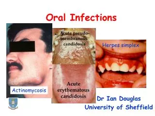
Necrotizing Ulcerative Gingivitis •Rare specific infectious gingival disease of young persons. •Fusobacterium nucleatum, Treponema vincentii, and probably other bacteria play an important role. •Predisposing factors are emotional stress, smoking, poor oral hygiene, local trauma, and HIV infection.
Clinical features • •The characteristic clinical feature is painful necrosis of the interdental papillae and the gingival margins, and the formation of craters covered with a gray pseudomembrane. • •Spontaneous gingival bleeding, halitosis, and intense salivation are common. • •Fever, malaise, and lymphadenopathy are less common. • •Rarely, the lesions may extend beyond the gingiva (necrotizing ulcerative stomatitis). • Treatment • •Systemic metronidazole . • Mechanical gingival treatment.
Noma •Noma, also known as cancrumoris and gangrenous stomatitis, is a devastating disease of malnourished children that is characterized by a destructive process of the orofacial tissues. •The condition is rare in developed countries. •Necrosis of tissue occurs as a consequence of invasion by anaerobic bacteria in a host whose systemic health is significantly compromised.
The reported predisposing factors include the following: ● Poverty ● Malnutrition or dehydration ● Poor oral hygiene ● Poor sanitation ● Unsafe drinking water ● Proximity to unkempt livestock ● Recent illness ● Malignancy ● An immunodeficiency disorder, including AIDS
Clinical Features. •It typically affects children. •The initial lesion of noma is a painful ulceration, rapidly and eventually becomes necrotic. •Denudation of involved bone may follow, eventually leading to necrosis and sequestration. •Teeth in the affected area may become loose and may exfoliate. •Penetration of organisms into the cheek, lip, or palate may also occur, resulting in necrotic lesions.
Treatment • •Therapy involves treating the underlying predisposing condition, as well as the infection itself. • Therefore fluids, electrolytes, and general nutrition are restored, along with the introduction of antibiotics
Syphilis • •Syphilis is a relatively common sexually transmitted disease Caused by Treponema pallidum. • •Syphilis may be acquired (common) or congenital (rare). • Acquired syphilis is classified as primary, secondary and tertiary.
Clinical features. • primary stage :- • Chancre that appears at the site of inoculation, usually three weeks after the infection. • Oral chancre appears in about 5–10% of cases, and clinically presents as a painless ulcer with a smooth surface, raised borders, and an indurated base. • •Regional lymphadenopathy is a constant finding
The secondary stage • Begins 6–8 weeks after the appearance of the chancre, and lasts for 2–10 weeks. • •Oral lesions are mucous patches (common), macular syphilids, and • condylomata lata (rare). • •Constitutional symptoms and signs (malaise, low-grade fever, headache, lacrimation, sore throat, weight loss, myalgias and multiple arthralgias, generalized lymphadenopathy). • As well as cutaneous manifestations (macular syphilids, papular syphilids, condylomata lata, nail involvement, hair loss, atypical rash, etc.) are constant findings.
Tertiary syphilis • Begins after a period of 4–7 years. • •Oral lesions are gumma, atrophic glossitis, and interstitial glossitis
Laboratory tests: • Dark-field microscopic examination, and serological tests. • Venereal Disease Research Laboratory (VDRL). • fluorescent treponemal antibody, absorbed (FTA-ABS). • Treponema pallidum immobilization (TPI). • rapid plasma reagin (RPR). • The most common oral lesions in congenital syphilis are a high-arched palate, short mandible, Hutchinson’s teeth, and Moon’s or mulberry molars.
Histopathology •The basic tissue response to T. pallidum infection consists of a proliferative endarteritis and infiltration of plasma cells. •Spirochetes can be demonstrated in the tissues of various lesions of syphilis using silver stains, although they may be scant in tertiary lesions. •Gummas may show necrosis and greater numbers of macrophages, resulting in a granulomatous lesion that is similar to other conditions, such as tuberculosis (TB) Treatment Penicillin is the antibiotic of choice. Erythromycin or cephalosporins are good alternatives
Tuberculosis Tuberculosis is a chronic, granulomatous, infectious disease that primarily affects the lungs, caused by Mycobacterium tuberculosis. .
Clinical features •The oral lesions are rare, and usually secondary to pulmonary tuberculosis. •The tuberculous ulcer is the most common feature. Clinically, the ulcer is painless and irregular, with a thin undermined border and a vegetating surface, usually covered by a gray-yellowish exudate.
•The surrounding tissues are inflamed and indurated. •The dorsum of the tongue is the most commonly affected site, followed by the lip, buccal mucosa, and palate. •Osteomyelitis of the jaws, periapical granuloma, regional lymphadenopathy, and scrofula are less common oral manifestations
Histopathology •Granulomatous inflammation, show central caseous necrosis. •In tissues, focal zones of macrophages become surrounded by lymphocytes and fibroblasts. •The macrophages develop an abundant eosinophilic cytoplasm, giving them a superficial resemblance to epithelial cells(epithelioid cells).
•Fusion of macrophages results in the appearance of Langerhans giant cells, in which nuclei are distributed around the periphery of the cytoplasm. •As the granulomas age, central necrosis occurs; this is usually referred to as caseous necrosis because of the gross cheesy texture of these zones. •A Ziehl-Neelsen or Acid fast stain must be used to confirm the presence of the organism in the granulomas.
Actinomycosis • •Chronic bacterial disease caused by Actinomyces israelii, an anaerobic , gram-positive bacterium. • •Infection appears after trauma, surgery, or previous infection. • Clinically • •Swelling of the mandible that may simulate a pyogenic infection. • •The clinical course ranges from acute to chronic. • •The skin lesions are indurated and are described as having a “woody hard” consistency. • Pus draining from the chronic lesion may contain small yellow granules, known as sulfur granules, which represent aggregates of A. israelii organisms. • •Radiographically, this infection presents as a lucency with irregular and ill-defined margins.
Histopathology •A granulomatous inflammatory response with central abscess formation is seen in actinomycosis. •At the center of the abscesses, distinctive colonies of gram-positive organisms may be seen. Radiating from the center of the colonies are numerous filaments with clubbed ends.