Oral Mucosa
Oral Mucosa. Dr Jamal Naim PhD in Orthodontics. Definition. Mucous membrane is used to describe the moist lining of the gastrointestinal tract, nasal passages and other cavities of the body that communicate with he exterior.

Oral Mucosa
E N D
Presentation Transcript
Oral Mucosa Dr Jamal Naim PhD in Orthodontics
Definition Mucous membrane is used to describe the moist lining of the gastrointestinal tract, nasal passages and other cavities of the body that communicate with he exterior. In the oral cavity this lining is called oral mucous membrane or oral mucosa. The oral mucosa is located between skin and gastrointestinal mucosa and shows some of the properties of each.
Definition • Like skin the oral mucosa consists of two tissue components: • A covering epithelium and • underlying connective tissue • Both of those tissues together perform a common function and so we consider them as an organ.
Functions of the oral mucosa • The main functions of the oral mucosa are: • Protection • Sensation • Secretion • Thermal regulation
Protection • OM separates and protects deeper tissues and organs from the environment of the oral cavity; • Protection from mechanical stress such as compression, stretching, shearing and abrasions. • Protection from chemical substances and toxins. • Protection from the pathologic effect of the microorganisms in the oral cavity.
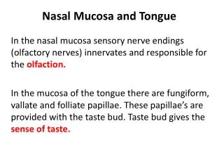
Sensation • The sensory function of the OM is important because it provide considerable information about events in the oral cavity: • Receptors respond to temperature, touch and pain • Taste buds on the tongue • Receptors respond to water taste and give signal about the satisfaction of thrist.
Secretion Salivary secretion by the salivary glands, major and minor, is important for moistening the OM. The minor salivary glands, which are distributed among the oral cavity, are responsible for about 80% of the salivary secretion of mucous saliva. The mucous saliva (mucin) is an important factor for the lubrication and moistening of the OM.
Thermal regulation Only in some animals the OM plays an important role in the regulation of the body temperature. In humans there isn't any influence of the OM by the regulation of the body temperature.
Anatomical considerations The oral cavity consists of the oral vestibule and the oral cavity proper. Laying between them is the alveolar process bearing the teeth. Hard and soft palate form the roof of the oral cavity proper. The floor of the mouth and the tongue form the inferior border of the oral cavity proper.
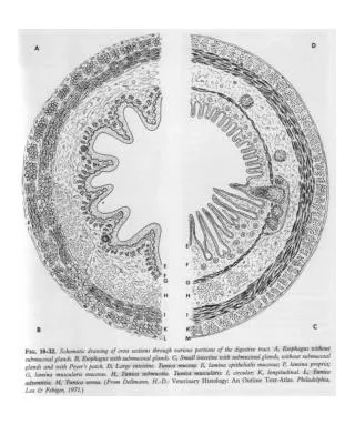
Anatomical considerations • There are three main types of the oral mucosa, identified according their primary function: • Masticatory mucosa • Lining mucosa • Specialized mucosa • The larger part of the OM is lining mucosa (60%), followed by the masticatory (25%) and specialized mucosa (15%)
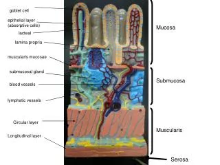
Components of The OM • The two main tissue components of the OM are: • The oral epithelium, it is a stratified squamous epithelium • The lamina propria, it is the underlying connective tissue. • The interface between oral epithelium and lamina propria (basement membrane) is usually irregular and the so called connective tissue papillae interdigitate with the epithelial ridges.
Basement membrane Connective tissue papillae
Components of The OM The tissue component under the oral mucosa is the submucosa and it is less easy to recognize the junction between them than between oral epithelium and lamina propria. In some regions such as cheek and lips a layer of loose fatty or glandular connective tissue containing the major blood vessels separates the OM from underlying bone or muscles.
Components of The OM The components of the submucosa determine the flexibility of the attachment of the oral mucosa to the underlying structures. For example we speak about mucoperiosteum in regions such as hard palate because the OM is attached directly to the palatal bone. Mucoperiosteum provides a firm inelastic attachment of the OM to bone.
Oral mucoperiosteum: it lacks on submucosa (e. g. region of the intermaxilary suture
Components of The OM In the OM there are minor salivary glands, sebaceous cells and nodules of lymphoid tissues. The largest accumulation of lymphoid tissue are found in the posterior part of oral cavity, where they form the lingual, palatine and pharyngeal tonsils. Those accumulations are known as the waldeyer's ring
Oral Epithelium • It is derived from the embryonic ectoderm and forms the primary barrier between oral environment and underlying tissues. • It consists of stratified squamous cells arranged in many layers (strata) • It maintains its continuity by a continuous cell renewal: • Cell proliferation • Cell maturation
Oral Epithelium • The cells of oral epithelium that take part in the renewal are: • Progenitor cells: the function is to divide and provide new cells • Maturing cells: they undergo a process of maturation to form the protective cell layer.
Epithelial proliferation The progenitor cells are located in the basement membrane in thin epithelia (floor of the mouth) and in the lower two to three cell layers in thicker epithelia (cheek) Progenitor cells divide into new progenitor cells or into maturing cells.
Epithelial proliferation The turnover time is the period of time is needed by a cell to divide and pass through the entire epithelium. The turnover time is 52 to 75 days in skin, 41 to 57 days in the gingiva and 25 days in the cheek.
Epithelial maturation • Cells that are driven from progenitor cells and ready for maturation passes the entire epithelium to form the protective layer. • In general maturation in the oral cavity follows two main patterns: • Keratinization • Nonkeratinization
Epithelial maturation Keratinization or cornification is the formation of a surface of keratin. Such process is seen in the oral mucosa of the palate, gingiva and in some regions of the tongue dorsum. A keratinized epithelium shows in histological sections a number of layers (strata):
Epithelial maturation • The basal cell layer - stratum basale - contains cuboidal or columnar cells and is adjacent to the basement membrane. • The cells of the st. basale shows the highest mitotic activity.
Epithelial maturation • Theprickle cell layer- stratum spinosum - lies above the first one and contains several rows of elliptical or spherical cells. • The cells have cytoplasmatic processes that have contact with other processes of other cells.
Epithelial maturation The mechanical adhesions between the cells are called desmosomes. Both, st. basale and st. spinosum, together constitute half to two third of the thickness of the oral epithelium.
Epithelial maturation • The granular layer - stratum granulosum - is the next layer and consists of larger flattened cells containing small granules.
Epithelial maturation • The keratinized layer - stratum corneum – is the surface layer and consists of flattened (squamous) cells. squamous cells. St. corneum
Epithelial maturation Nonkeratinization occurs in regions with less mechanical influences to the OM, such as cheek, lips, underside of the tongue and the soft palate. Nonkeratinized epithelium is usually thicker than keratinized epithelium. No sudden changes in the cells above the st. spinosum occur in nonkeratinized epithelium, and the outer half of the tissue is divided into two zones:
Epithelial maturation • Intermediate layer - stratum intermedium - • Superficial layer – stratum superficiale –
Epithelial maturation in Non-keratinized oral mucosa Epithelial maturation in keratinized oral mucosa
Epithelial maturation In so called parakeratinized mucosa, such as parts of the hard palate and the gingiva, in the surface layer the nuclei are shrunken and retained in many or all squames. Also keratohyaline granules are present in this layer. Such phenomenon is a normal event in the oral epithelium, but not true for the epidermis, where parakeratinization is associated with diseases such as psoriasis.
Ultrastructure in the OE In the next session