Mucosa
780 likes | 923 Vues
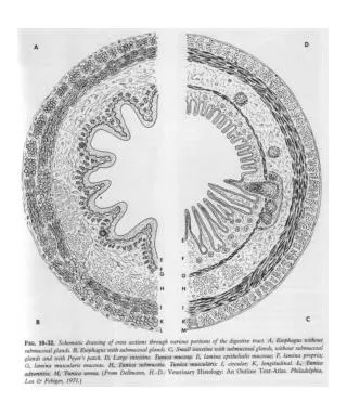
Throughout the remainder of the digestive system, the histological composition of the alimentary canal can be described by the following four layers. Mucosa

Mucosa
E N D
Presentation Transcript
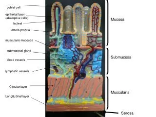
Throughout the remainder of the digestive system, the histological composition of the alimentary canal can be described by the following four layers.
Mucosa • The lumen is lined by an epithelium, which rests on a vascular c.t. the lamina propria,which contains bl. vessels, lymph nodules and glands. The lamina propria is surrounded by a band of smooth muscle(muscularis mucosae). These three tissues are collectively referred to as the mucosaof thealimentary canal .
Submucosa :Is a connective tissue containing blood vessels, lymph nodules,the submucosal (Meissener's) nerve plexus and submucosal glands (oesophagus and duodenum).
Muscularis Externa: Inner circular layer and outer longitudinal. Between these two layers is the myenteric (Auerbach) nerve plexus
Adventitia/Serosa: The alimentary canal is surrounded by a layer of loose connective tissue, the adventitia. In the case of the intraperitoneal parts of the alimentary canal, a simple squamous epithelium to a layer of loose of connective tissue, forms the serosa.
The Oesophagus • In the oesophagus the mucosa is formed by a stratified squamous epithelium (non-keratinised) and a well-defined lamina propria and muscularis mucosae. • Oesophageal glands are located in the submucosa.
The muscularis externa contains striated muscle in its upper onethird, a mixture of striated muscle and smooth muscle in its middle one-third and smooth muscle in its lower one-third.
The adventitia surrounds the intrathoracic part which consists only of a layer of loose connective tissue. A serosa forms the outermost part of the short intraperitoneal segment of the oesophagus.
GASTROINTESTINAL TRACT • The gastrointestinal tract (GIT) comprises the stomach, duodenum, jejunum, ileum, colon, rectumand anal canal. The GIT and oesophagus form the alimentary canal.
The Stomach • Anatomically, the stomach is divided into :a cardiac part, fundus, body or corpus,anda pyloric part (pyloric antrum and pyloric canal) The Mucosa (epithelium, lamina propria, muscularis mucosae).The mucosa is thrown into longitudinal folds (gastric folds or rugae), which disappear when the stomach is fully distended.
On the mucosal surface we see small, depressions (gastricpits). Simple, branched tubular gastric glands open into the bottom of the gastric pits. The structure and cellular composition of the surface epithelium which is simple, tall columnar, does not change throughout the stomach. These cells secrete mucus.
Cardiac glands: Cardiac glands are branched tubular glands which contain mainly mucus-producing cells. • Principal (or corpus-fundic) glands Each glandular tubule consists of : a deep body, and neck .In principal glands we find four cell types: chief cells, parietal cells, mucous neck cells and endocrine cells.
Chief cells (or zymogenic cells) are the most numerous of the four types. They occur primarily in the body(base) of the glands. They produce pepsinogen. Parietal cells (or oxyntic cells) occur in the neck of the glands. Parietal cell secrete HCL and intrinsic factor, which is necessary for the resorption of vitamin B12.
Mucous neck cells are found between the parietal cells in the neck of the gland.Endocrine cells are part of the gastro-entero-pancreatic (GEP) endocrine system. The endocrine cells in the gastric mucosa are gastrin-producing cells (G cells) and somatostatin-producing cells (D cells).
Pyloric glands Pyloric glands are more coiled than principal glands.Gastrin-producing cells, are more frequent than in principal glands. A few parietal cells may be present. • The lamina propria is formed by a very cell-rich loose c.t. (fibroblasts, lymphocytes, plasma cells, macrophages, eosinophilic leukocytes and mast cells).
The muscularis mucosae of the stomach contains both circular and longitudinal layers of muscle cells. • Large blood vessels, lymph vessels and nerves and the submucosal (Meissener's) nerve plexus are located in the submucosa which consists of loose connective tissue.
The muscularis externa consists of three layers of muscles: an inner oblique layer, a middle circular layer and an outer longitudinal layer. • Serosa: is connective tissue and mesothelium.
Small Intestine • The small intestine is divided into duodenum, jejunum and ileum. The three segments have the same basic histological organization.
The mucosa of the small intestine has variousstructural features which increase theluminal surface area. • Plicaecirculares(of Kerkering) are macroscopically visible, crescent-shaped folds of the mucosa and submucosa. Plicae circulares are permanent structures
The entire intestinal mucosa forms intestinalvilli(elevations of epithelium and lamina propria).The surface of the villi is formed by a simple columnar epithelium. Each absorptive cell, or enterocyte, of the epithelium forms numerous microvilli.
Between the intestinal villi we see the openings of simple tubular glands, the crypts of Lieberkühn. They extend through the lamina propria down to the muscularis mucosae. Undifferentiated cells close to the bottom of the crypts regenerate the epithelium.
Other epithelial cells in the crypts correspond largely to those in the epithelium of the intestinal villi.Paneth cell release a number of antibacterial substances and goblet cells secrete mucus. • The lamina propria is, similar to the lamina propria of the stomach.
Lymph nodules may form aggregations of 30-50 nodules in the lamina propria of the ileum. These large aggregations are called Peyer's patches.
The muscularis mucosae has two layers and extends into the intestinal villi, where the smooth muscle cells form a longitudinal bundle in the centre of the villi .Blood vessels and lymphatic capillaries(lacteals) are also present
The Submucosa The submucosa contains glands only in theduodenum. Submucosal glands of the duodenum are also called Brunner's glands. Their secretion is mucous and slightly alkaline and protects the duodenal mucosa.
The large intestine constitutes the terminal part of the digestive system. It is divided into three main sections: cecum including the appendix, colon, andrectum with the anal canal. • The surface of the mucosa is smooth as there are no plicae circularesor intestinal villi.
Crypts of Lieberkühn are present. Goblet cells are more than in the small intestine. • There is lamina propria between the glands. The muscularis mucosae forms two layers. The appearance of the muscularis externa is different from that of the small intestine.
The inner circular layer of muscle forms the usual sheath around the large intestine, but the outer longitudinal muscle layer forms three flattened strands, the taeniae coli. • The adventitia forms small pouches (appendices epiploicae) filled with fatty tissue along the large intestine.
Specialized Sections of the Large Intestine • The vermiform appendix:The most important of its features is the thickening of its walls primarily due to large accumulations of lymphoid nodules in the lamina propria.