Development of an Optimized Scanning X-Ray Microdiffraction System
Development of an Optimized Scanning X-Ray Microdiffraction System for Materials Research and Education Ersan Ustundag, Iowa State University, DMR IMR- 0416243.

Development of an Optimized Scanning X-Ray Microdiffraction System
E N D
Presentation Transcript
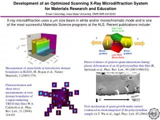
Development of an Optimized Scanning X-Ray Microdiffraction System for Materials Research and Education Ersan Ustundag, Iowa State University, DMR IMR-0416243 Direct evidence of grain-to-grain interactions during plastic deformation of an Al polycrystalline thin film (R. Spolenak et al., Phys. Rev. Lett., 90 (2003) 096102) Measurement of strain fields at ferroelectric domain boundaries in BaTiO3(R. Rogan et al., Nature Materials, 2 (2003) 379) Characterization and shear stress measurements at twin domain boundaries of a superconducting YBCO thin film (W.A. Caldwell et al., Phys. Rev. Lett., 21 (2004), 216105 New mechanism of grain growth under current evidenced in electromigrated b-Sn polycrystalline sample (A.T. Wu et al., Appl. Phys. Lett. 85 (2004) 2490) X-ray microdiffraction uses a m size beam in white and/or monochromatic mode and is one of the most successful Materials Science programs at the ALS. Recent publications include:
Development of an Optimized Scanning X-Ray Microdiffraction System for Materials Research and Education Ersan Ustundag, Iowa State University, DMR IMR-0416243 Diffraction pattern from a single CuNW nanowire (100 nm diameter, 10 mm long). Successful NSF DMR-IMR grant led to the commissioning of a dedicated X-ray microdiffraction beamline at the ALS with enhanced capabilities Enhanced capabilities will enable new science: Higher flux and smaller X-ray spot size (~100 nm): looking at the mechanical properties of nanomaterials Broader spectrum range yields increased strain/stress sensitivity • Front-end commissioning underway (completion in October 2005) • Installation of the end-station at new beamline programmed for Spring 2006 Broader spectrum range and special fixtures will allow 3D microdiffraction to probe properties at granular level in bulk materials