Download
1 / 112
1.13k likes | 1.63k Vues
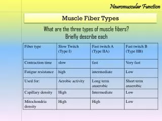
Neuromuscular Fundamentals. Anatomy and Physiology of Human Movement 420:050. Outline. Introduction Structure and Function Fiber Arrangement Muscle Actions Role of Muscles Neural Control Factors that Affect Muscle Tension. Introduction.

E N D
Neuromuscular Fundamentals Anatomy and Physiology of Human Movement 420:050
Outline • Introduction • Structure and Function • Fiber Arrangement • Muscle Actions • Role of Muscles • Neural Control • Factors that Affect Muscle Tension
Introduction • Responsible for movement of body and all of its joints • Muscles also provide • Protection • Posture and support • Produce a major portion of total body heat • Over 600 skeletal muscles comprise approximately 40 to 50% of body weight • 215 pairs of skeletal muscles usually work in cooperation with each other to perform opposite actions at the joints which they cross • Aggregate muscle action - muscles work in groups rather than independently to achieve a given joint motion
Muscle Tissue Properties • Irritability or Excitability - property of muscle being sensitive or responsive to chemical, electrical, or mechanical stimuli • Contractility - ability of muscle to contract & develop tension or internal force against resistance when stimulated • Extensibility - ability of muscle to be passively stretched beyond it normal resting length • Elasticity - ability of muscle to return to its original length following stretching
Outline • Introduction • Structure and Function • Fiber Arrangement • Muscle Actions • Role of Muscles • Neural Control • Factors that Affect Muscle Tension
Structure and Function • Nervous system structure • Muscular system structure • Neuromuscular function
Figure 14.1, Marieb & Mallett (2003). Human Anatomy. Benjamin Cummings.
Nervous System Structure • Integration of information from millions of sensory neurons action via motor neurons Figure 12.1, Marieb & Mallett (2003). Human Anatomy. Benjamin Cummings.
Nervous System Structure • Organization • Brain • Spinal cord • Nerves • Fascicles • Neurons Figure 12.2, Marieb & Mallett (2003). Human Anatomy. Benjamin Cummings. Figure 12.7, Marieb & Mallett (2003). Human Anatomy. Benjamin Cummings.
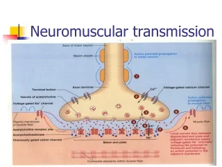
Nervous System Structure • Both sensory and motor neurons in nerves Figure 12.11, Marieb & Mallett (2003). Human Anatomy. Benjamin Cummings.
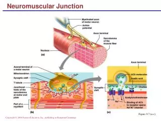
Nervous System Structure • The neuron: Functional unit of nervous tissue (brain, spinal cord, nerves) • Dendrites: Receptor sites • Cell body: Integration • Axon: Transmission • Myelin sheath: Protection and speed • Nodes of Ranvier: Saltatory conduction • Terminal branches: Increased innervation • Axon terminals: Connection with muscular system • Synaptic vescicles: Delivery mechanism of “message” • Neurotransmitter: The message
Dendrites Cell body Axon Myelin sheath Node of Ranvier Terminal ending Terminal branch Figure 12.4, Marieb & Mallett (2003). Human Anatomy. Benjamin Cummings.
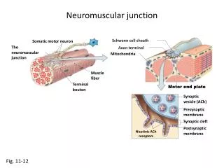
Figure 12.8, Marieb & Mallett (2003). Human Anatomy. Benjamin Cummings. Terminal ending Synaptic vescicle Neurotransmitter: Acetylcholine (ACh)
Figure 12.19, Marieb & Mallett (2003). Human Anatomy. Benjamin Cummings.
Structure and Function • Nervous system structure • Muscular system structure • Neuromuscular function
Classification of Muscle Tissue • Three types: 1. Smooth muscle 2. Cardiac muscle 3. Skeletal muscle
Skeletal Muscle: Properties • Extensibility: The ability to lengthen • Contractility: The ability to shorten • Elasticity: The ability to return to original length • Irritability: The ability to receive and respond to stimulus
Muscular System Structure • Organization: • Muscle (epimyseum) • Fascicle (perimyseum) • Muscle fiber (endomyseum) • Myofibril • Myofilament • Actin and myosin • Other Significant Structures: • Sarcolemma • Transverse tubule • Sarcoplasmic reticulum • Tropomyosin • Troponin
Figure 10.1, Marieb & Mallett (2003). Human Anatomy. Benjamin Cummings.
Figure 10.4, Marieb & Mallett (2003). Human Anatomy. Benjamin Cummings.
http://staff.fcps.net/cverdecc/Adv%20A&P/Notes/Muscle%20Unit/sliding%20filament%20theory/slidin16.jpghttp://staff.fcps.net/cverdecc/Adv%20A&P/Notes/Muscle%20Unit/sliding%20filament%20theory/slidin16.jpg
Figure 10.8, Marieb & Mallett (2003). Human Anatomy. Benjamin Cummings.
Structure and Function • Nervous system structure • Muscular system structure • Neuromuscular function
Neuromuscular Function • Basic Progression: 1. Nerve impulse 2. Neurotransmitter release 3. Action potential along sarcolemma 4. Calcium release 5. Coupling of actin and myosin 6. Sliding filaments
Nerve Impulse • What is a nerve impulse? -Transmitted electrical charge -Excites or inhibits an action -An impulse that travels along an axon is an ACTION POTENTIAL
Nerve Impulse • How does a neuron send an impulse? -Adequate stimulus from dendrite -Depolarization of the resting membrane potential -Repolarization of the resting membrane potential -Propagation
Nerve Impulse • What is the resting membrane potential? -Difference in charge between inside/outside of the neuron -70 mV Figure 12.9, Marieb & Mallett (2003). Human Anatomy. Benjamin Cummings.
Nerve Impulse • What is depolarization? -Reversal of the RMP from –70 mV to +30mV Propagation of the action potential Figure 12.9, Marieb & Mallett (2003). Human Anatomy. Benjamin Cummings.
Nerve Impulse • What is repolarization? -Return of the RMP to –70 mV Figure 12.9, Marieb & Mallett (2003). Human Anatomy. Benjamin Cummings.
+30 mV -70 mV
Neuromuscular Function • Basic Progression: 1. Nerve impulse 2. Neurotransmitter release 3. Action potential along sarcolemma 4. Calcium release 5. Coupling of actin and myosin 6. Sliding filaments
Release of the Neurotransmitter • Action potential axon terminals 1. Calcium uptake 2. Release of synaptic vescicles (ACh) 3. Vescicles release ACh 4. ACh binds sarcolemma
Figure 12.8, Marieb & Mallett (2003). Human Anatomy. Benjamin Cummings. Ca2+ ACh
Figure 14.5, Marieb & Mallett (2003). Human Anatomy. Benjamin Cummings.
Neuromuscular Function 1. Nerve impulse 2. Neurotransmitter release 3. Action potential along sarcolemma 4. Calcium release 5. Coupling of actin and myosin 6. Sliding filaments
AP Along the Sarcolemma • Action potential Transverse tubules 1. T-tubules carry AP inside 2. AP activates sarcoplasmic reticulum
Figure 14.5, Marieb & Mallett (2003). Human Anatomy. Benjamin Cummings.
Neuromuscular Function 1. Nerve impulse 2. Neurotransmitter release 3. Action potential along sarcolemma 4. Calcium release 5. Coupling of actin and myosin 6. Sliding Filaments
Calcium Release • AP T-tubules Sarcoplasmic reticulum 1. Activation of SR 2. Calcium released into sarcoplasm
CALCIUM RELEASE Sarcolemma
Neuromuscular Function 1. Nerve impulse 2. Neurotransmitter release 3. Action potential along sarcolemma 4. Calcium release 5. Coupling of actin and myosin 6. Sliding filaments
Coupling of Actin and Myosin • Tropomyosin • Troponin
Blocked Coupling of actin and myosin
Neuromuscular Function 1. Nerve impulse 2. Neurotransmitter release 3. Action potential along sarcolemma 4. Calcium release 5. Coupling of actin and myosin 6. Sliding filaments
Sliding Filament Theory • Basic Progression of Events 1. Cross-bridge 2. Power stroke 3. Dissociation 4. Reactivation of myosin
Cross-Bridge • Activation of myosin via ATP -ATP ADP + Pi + Energy -Activation “cocked” position
Power Stroke • ADP + Pi are released • Configurational change • Actin and myosin slide
Dissociation • New ATP binds to myosin • Dissociation occurs
Reactivation of Myosin Head • ATP ADP + Pi + Energy • Reactivates the myosin head • Process starts over • Process continues until: -Nerve impulse stops -AP stops -Calcium pumped back into SR -Tropomyosin/troponin back to original position