Download
1 / 69
700 likes | 975 Vues
Chapter 2 Neuromuscular Fundamentals. Skeletal Muscles. Responsible for movement of body and all of its joints Muscle contraction produces force that causes joint movement Muscles also provide protection posture and support produce a major portion of total body heat. Muscle Nomenclature.

E N D
Skeletal Muscles • Responsible for movement of body and all of its joints • Muscle contraction produces force that causes joint movement • Muscles also provide • protection • posture and support • produce a major portion of total body heat
Muscle Nomenclature • Muscles are usually named due to • visual appearance • anatomical location • function • Shape – deltoid, rhomboid • Size – gluteus maximus, teres minor • Number of divisions – triceps brachii • Direction of its fibers – external abdominal oblique
Shape of Muscles & Fiber Arrangement • Cross section diameter • factor in muscle’s ability to exert force • greater cross section diameter = greater force exertion • Muscle’s ability to shorten • longer muscles can shorten through a greater range • more effective in moving joints through large ranges of motion Neuromuscular Fundamentals
Muscle Terminology • Origin • Structurally, the proximal attachment of a muscle or the part that attaches closest to the midline or center of the body • Functionally & historically, the least movable part or attachment of the muscle • Insertion • Structurally, the distal attachment or the part that attaches farthest from the midline or center of the body • Functionally & historically, the most movable part is generally considered the insertion
Types of muscle contraction • Muscle contractions can be used to cause, control, or prevent joint movement or • to initiate or accelerate movement of a body segment • to slow down or decelerate movement of a body segment • to prevent movement of a body segment by external forces • All muscle contractions are either isometric or isotonic Neuromuscular Fundamentals
Isometric Isotonic Concentric Eccentric Types of muscle contraction Muscle Contraction (under tension) Preventing Motion Neuromuscular Fundamentals
Types of muscle contraction • Isometric contraction • active tension is developed within muscle but joint angles remain constant • static contractions • significant amount of tension may be developed in muscle to maintain joint angle in relatively static or stable position • may be used to prevent a body segment from being moved by external forces • preventing motion Neuromuscular Fundamentals
Types of muscle contraction • Concentric contractions involve muscle developing active tension as it shortens • Causing motion • Eccentric contractions involvethe muscle lengthening under active tension • Controlling motion
Role of Muscles • Agonist muscles • cause joint motion through a specified plane of motion when contracting concentrically • known as primary or prime movers • Some agonist muscles, because of their relative location, size, length, or force generation capacity, are able to contribute significantly more to the joint movement than other agonists • Assisters or assistant movers • Agonist muscles that contribute significantly less to the joint motion
Role of Muscles • Antagonistmuscles • located on opposite side of a joint from the agonist • have the opposite concentric action • known as contralateral muscles • work in cooperation with agonist muscles by relaxing & allowing movement • when contracting concentrically perform the opposite joint motion of agonist • Ex. quadriceps muscles are antagonists to hamstrings in knee flexion
Role of Muscles • Stabilizers • surround joint or body part • contract to fixate or stabilize the area to enable another limb or body segment to exert force & move • known as fixators • essential in establishing a relatively firm base for the more distal joints to work from when carrying out movements • Ex. biceps curl • muscles of scapula & glenohumeral joint must contract in order to maintain shoulder complex & humerus in a relatively static position so that the biceps brachii can more effectively perform curls • Proximal stability is needed for distal mobility.
Role of Muscles • Synergist • assist in action of agonists • not necessarily prime movers for the action • known as guiding muscles • assist in refined movement & rule out undesired motions
Role of Muscles • Force Couples • Force couples occur when two or more forces are pulling in different directions on an object, causing the object to rotate about its axis • Coupling of muscular forces together in the body can result in a more efficient movement
Lines of Pull Consider the following • Exact locations of bony landmarks to which muscles attach proximally & distally and their relationship to joints • Planes of motion through which a joint is capable of moving • Muscle’s relationship or line of pull relative to the joint’s axes of rotation • As a joint moves the line of pull may change & result in muscle having a different or opposite action than in the original position • Effect of a muscle’s relative length on its ability to generate force
Factors affecting muscle tension development • All or None Principle - regardless of number, individual muscle fibers within a given motor unit will either fire & contract maximally or not at all • Difference between lifting a minimal vs. maximal resistance is the number of muscle fibers recruited • The number of muscle fibers recruited may be increased by • activating those motor units containing a greater number of muscle fibers • activating more motor units • increasing the frequency of motor unit activation
Motor Control • A motor neuron and the muscle fibers it innervates. • Fine control • small motor units contain as few as 20 muscle fibers per nerve fiber • eye muscles • Allow for greater dexterity because of a lower neuron to muscle fiber ratio. • Gross control • gastrocnemius muscle has 1000 fibers per nerve fiber • One motor neuron controls many muscle fibers which allows for strength production.
Recruitment and Stimulus Intensity • Strength of muscle contraction is dependant of # of motor units recruited • Multiple motor unit summation • The harder the activity is the more motor units will be recruited. • Lifting 1 lb vs. 100 lbs • How do you explain the rapid strength gains when you first start training?
All or None Principle • Typical muscle contraction • The number of motor units responding (and number of muscle fibers contracting) within the muscle may vary significantly from relatively few to virtually all • depending on the number of muscle fibers within each activated motor unit & the number of motor units activated
Metabolism and Skeletal Muscle Fibers Types • There are 3 different types skeletal muscle fibers based histological differences, duration of a twitch and the method of ATP production • slow oxidative fibers • fast oxidative fibers • fast glycolytic fibers • Proportions genetically determined
Fast Glycolytic, Fast-Twitch Fibers • Fast glycolytic, fast-twitch fibers: • rich in enzymes for phosphagen and glycogen-lactic acid systems • Limited # of mitochondria and high concentration of glycogen stores makes it adapted for anaerobic metabolism • a lack of myoglobin in glycolytic fibers results in a white color • sarcoplasmic reticulum releases calcium quickly so contractions are quicker which are required for movements that produce speed and power. • extraocular eye muscles, gastrocnemius and biceps brachii
Slow- Twitch Fibers • Slow oxidative, slow-twitch fibers • Oxidative fibers contain greater amounts of mitochondria and myoglobin which binds oxygen. • Rich blood supply and high concentration of myoglobin these fibers appear red in color. • adapted for endurance (resistant to fatigue) • Soleus and postural muscles of the back are predominantly this type.
Fast Oxidative Fibers • Fast oxidative fibers: • characteristics of both fast and slow fibers. • have a fast twitch (use ATP quickly) • Increased mitochondria make it moderately resistant to fatigue • Usually make up 10% of fibers. • Training will make these fibers adapt to become functionally more fast or slow.
Cellular Adaptations to Physical Demands • Strength training: high intensity training stresses anaerobic pathways. • Increased # and size of glycolytic associated enzymes and substrates • ATP, creatine phosphate and glycogen • Endurance training: Enhance the aerobic pathways. • increased # and size of mitochondrial membranes and associated enzymes. • This will increase O2 uptake ( VO2 max) which will delay the formation of lactic acid.
Factors affecting muscle tension development • Number of muscle fibers per motor unit varies significantly • From less than 10 in muscles requiring precise and detailed such as muscles of the eye • To as many as a few thousand in large muscles that perform less complex activities such as the quadriceps Neuromuscular Fundamentals
Muscle Length - Tension Relationship • Generally, depending upon muscle involved • Greatest amount of tension can be developed when a muscle is stretched between 100% to 130% of its resting length • Stretch beyond 100% to 130% of resting length significantly decreases the amount of force production • A proportional decrease in ability to develop tension occurs as a muscle is shortened • When shortened to around 50% to 60% of resting length ability to develop contractile tension is essentially reduced to zero
Muscle Length - Tension Relationship • Maximal ability of a muscle to develop tension & exert force varies depending upon the length of the muscle during contraction Neuromuscular Fundamentals
Muscle Length - Tension Relationship • Ex. 1 Increasing ability to exert force • squat slightly to stretch the gastroc, hamstrings, & quadriceps before contracting same muscles concentrically to jump • Ex. 2. Reducing ability to exert force • isolate the gluteus maximus by maximally shortening the hamstrings with knee flexion Neuromuscular Fundamentals
Reciprocal Inhibition or Innervation • Antagonist muscles groups must relax & lengthen when the agonist muscle group contracts • This reciprocal innervation effect occurs through reciprocal inhibition of the antagonists • Activation of the motor units of the agonists causes a reciprocal neural inhibition of the motor units of the antagonists • This reduction in neural activity of the antagonists allows them to subsequently lengthen under less tension • Biceps contracts triceps must relax
Active & Passive Insufficiency • As muscle shortens its ability to exert force diminishes • Active insufficiency is reached when the muscle becomes shortened to the point that it can not generate or maintain active tension • Passively insufficiency is reached when the opposing muscle becomes stretched to the point where it can no longer lengthen & allow movement
Active Insufficiency • When a multi-joint muscle contracts simultaneously over 2 joints the muscle is unable to generate a maximum muscle contraction. • Ex. Rectus femoruscontracts concentrically to both flex the hip & extend the knee • Hamstrings: contracts both knee flexion and hip extension • Gastrocnemius: Knee flexion and plantar flexion • Biceps brachii: shoulder flexion and elbow flexion
Passive Insufficiency • Agonist muscle unable to reach full range of motion because of inadequate length of antagonistic two joint muscle. • Hamstrings can’t stretch enough to allow both maximal hip flexion & maximal knee extension. • Knee flexion cannot be fully achieved if the hip is fully extended. Rectus femoris length limits motion. • Finger flexion is limited if wrist flexion occurs simultaneously.
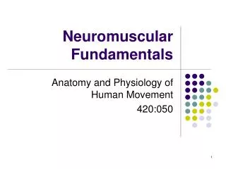
Proprioception • Subconscious mechanism by which body is able to regulate posture & movement by responding to stimuli originating in proprioceptors of the joints, tendons, muscles, & inner ear • Proprioceptors - internal receptors located in skin, joints, muscles, & tendons which provide feedback relative to: • tension, length, & contraction state of muscle. • position of body & limbs. • movements of joints. Neuromuscular Fundamentals
Proprioception • Proprioceptors specific to muscles • Muscles spindles • concentrated primarily in muscle belly between the fibers • sensitive to stretch & rate of stretch • Insert into connective tissue within muscle & run parallel with muscle fibers • Golgi tendon organs (GTO) • found serially in the tendon close to muscle tendon junction • sensitive to both muscle tension & active contraction • much less sensitive to stretch than muscle spindles • require a greater stretch to be activated
Muscle spindle • Muscle spindles & myotatic or stretch reflex • Rapid muscle stretch occurs • Afferent Impulse is sent to the CNS • CNS activates motor neurons of agonist muscle while inhibiting the antagonist muscle. • Stretched or agonistic muscle contracts
Monosynaptic Reflex • Ex. Knee jerk or patella tendon reflex Neuromuscular Fundamentals
Golgi Tendon Organ • Golgi tendon organ • Tension in tendons & GTO increases as muscle contract, which activates GTO • GTO stretch threshold is reached • Impulse is sent to CNS • CNS causes muscle to relax • facilitates activation of antagonists as a protective mechanism • GTO protects us from an excessive contraction by causing its muscle to relax
Chapter 3Basic Biomechanical Factors & Concepts Basic Biomechanical Factors & Concepts
Biomechanics • Biomechanics - study of the mechanics as it relates to the functional and anatomical analysis of biological systems • Necessary to study the body’s mechanical characteristics & principles to understand its movements • Mechanics - study of physical actions of forces • Musculoskeletal system may be thought of as a series of simple machines • Machines - used to increase mechanical advantage • Consider mechanical aspect of each component in analysis with respect to components’ machine-like function
Levers • Humans moves through a system of levers • Levers cannot be changed, but they can be utilized more efficiently • lever - a rigid bar that turns about an axis of rotation or a fulcrum • axis - point of rotation about which lever moves • Levers rotate about an axis as a result of force (effort, E) being applied to cause its movement against a resistance or weight • In the body • bones represent the bars • joints are the axes • muscles contract to apply force
Levers • Three points determine type of lever & for which kind of motion it is best suited • Axis (A)- fulcrum - the point of rotation • Point (F) of force application (usually muscle insertion) - effort • Point (R) of resistance application (center of gravity of lever) or (location of an external resistance) Basic Biomechanical Factors & Concepts
Levers Systems • 1st class lever – axis (A) somewhere between force (F) & resistance (R) • 2nd class lever – resistance (R) somewhere between axis (A) & force (F) • 3rd class lever – force (F) somewhere between axis (A) & resistance (R)
Levers • FAR1st | Force Arm| | Resistance Arm | F R A • ARF2nd | Resistance Arm | | Force Arm | R F A | Force Arm | • AFR3rd | Resistance Arm | F R A Basic Biomechanical Factors & Concepts
First-class Levers • Produce balanced movements when axis is midway between force & resistance (e.g., seesaw or scissors) • Produce speed & range of motion when axis is close to force, (triceps in elbow extension) • Produce force motion when axis is close to resistance (crowbar)
First-class Levers • Head balanced on neck in flexing/extending • Elbow extension in triceps applying force to olecranon (F) in extending the non-supported forearm (R) at the elbow (A) • Agonist & antagonist muscle groups are contracting simultaneously on either side of a joint axis • agonist produces force while antagonist supplies resistance
First-class Levers • Force is applied where muscle inserts in bone, not in belly of muscle • Ex. in elbow extension with shoulder fully flexed & arm beside the ear, the triceps applies force to the olecranon of ulna behind the axis of elbow joint • As the applied force exceeds the amount of forearm resistance, the elbow extends
Levers • The mechanical advantage of levers may be determined using the following equations: Mechanical advantage = ResistanceForce or Mechanical advantage = Length of force armLength of resistance arm
Torque and length of lever arms First class levers A, If the force arm & resistance arm are equal in length, a force equal to the resistance is required to balance it; B, As the force arm becomes longer, a decreasing amount of force is required to move a relatively larger resistance; C, As the force arm becomes shorter, an increasing amount of force is required to move a relatively smaller resistance
Second-class Levers • Produces force movements, since a large resistance can be moved by a relatively small force • Wheelbarrow • Nutcracker • Loosening a lug nut • Raising the body up on the toes
Second-class Levers • Plantar flexion of foot to raise the body up on the toes where ball (A) of the foot serves as the axis as ankle plantar flexors apply force to the calcaneus (F) to lift the resistance of the body at the tibial articulation (R) with the foot • Relatively few 2nd class levers in body