Understanding Atrioventricular Canal and Inter-Atrial Septum Formation in Fetal Development
This document explores the intricate process of atrioventricular canal formation and the development of the inter-atrial septum. It details how the septum primum originates from the roof of the common atrium, leading to the formation of the ostium primum, and subsequent steps involving the ostium secundum and septum secundum. Additionally, it discusses the three main physiological fetal shunts: the Ductus Arteriosus, Foramen Ovalis, and Ductus Venosus, along with their locations, functions, and closure mechanisms postnatally.

Understanding Atrioventricular Canal and Inter-Atrial Septum Formation in Fetal Development
E N D
Presentation Transcript
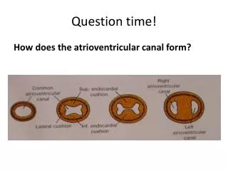
Question time! How does the atrioventricular canal form?
A B D C
D A B C
ESA Q: Describe the formation of the inter-atrial septum. (4 marks) • Septum primum forms from the roof of the common atrium towards the endocardial cushions and forms ostiumprimum • Ostiumsecundum forms higher up in septum primum and ostiumprimum closes • Septum secundum forms from the roof (to the right of septum primum) and forms the foramen ovalis.
What are the three physiological fetal shunts, where are they, what do they bypass, how do they close and what do they become?
Functional closure Anatomical closure LigamentumArteriosum Pulmonary A - Aorta DuctusArteriosus Lungs Foramen Ovalis Pressure Of LA increases. RV+ Lungs RA - LA Fossa Ovalis DuctusVenosus Liver Ligamentumvenosum Portal Vein - IVC