The Cardiovascular System
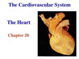
The Cardiovascular System. CONDUCTION SYSTEM. Activity 1. Draw and label in as much detail as possible the chambers of the heart and surrounding structures. Activity 1. Now complete the following diagram on the worksheet provided. Activity 2. We will now add more detail to the


The Cardiovascular System
E N D
Presentation Transcript
The Cardiovascular System CONDUCTION SYSTEM
Activity 1 Draw and label in as much detail as possible the chambers of the heart and surrounding structures.
Activity 1 Now complete the following diagram on the worksheet provided
Activity 2 We will now add more detail to the heart this information is known as the ‘Conduction System’
How the Heart Works • The heart works by producing impulses which spread and innervate the specialised muscle fibres. • Unlike skeletal muscle, the heart produces its own impulses (it is myogenic), and it is the conduction system of the heart which spreads the impulses and enables the heart to contract. http://www.youtube.com/watch?v=te_SY3MeWys&feature=related
How the Heart Works • The electrical impulse begins at the pacemaker: a mass of cardiac cells known as the sino-atrial node (S.A. node). • It is the rate at which the S.A. node emits impulses, that determines heart rate. • As the impulse is emitted, it spreads to adjacent interconnecting fibres of the atrium, which causes the atria to contract. • It then passes to another specialised mass of cells called the atrioventricular node (A.V. node).
How the Heart Works • The A.V. node acts as a distributor and passes the action potential to the Bundle of His, which, together with the branching Purkinje fibres, spread the excitation throughout the ventricles. • There is a delay of about 0.1 second from the time when the A.V. node receives stimulation, to when it distributes action potential through the ventricles.
How the Heart Works • This is crucial to allow completion of atrial contraction, before ventricular systole begins. • The relationship between the electrical activity of the heart and the cardiac cycle can be shown in an electrocardiogram (ECG)
Summary • Heart generates own electrical impulses, • At the sino-atrial node (pacemaker), • Impulse spreads through cardiac tissue in the atria, • This causes contraction of the atria, • The impulse carries on to Atrio-ventricular node, • The action potential moves into Bundle of His and spreads throughout the Purkinje fibres causing the ventricle to contract.
Introduction to the Cardiac Cycle • It is the process of cardiac contraction and blood transportation through the heart. • We are going to explain the sequence of events that takes place during one complete heartbeat. • Including filling and emptying of the blood into the arterial system.
Four Stage to Each Beat What does all this really mean? 1) Atrial Diastole Diastole means that heart is filling 2) Ventricular Diastole 3) Atrial Systole Systole means that the heart is emptying 4) Ventricular Systole
Atrial Diastole • Takes the 0.7 seconds. • The upper chamber of the heart are filled with blood returning from; • The body via the venaecavae to the right atrium. • The lungs via the pulmonary vein to the left atrium. • At this time the AV valves are shut but as the atria fill with blood, atrial pressure overcomes ventricular pressure. • Since blood always moves from areas of high pressure to areas of low pressure, the atrioventricular values are forced open and ventricular diastole now takes place.
Ventricular Diastole • 0.5 seconds • During this stage the ventricles fill with blood and the semi-lunar valves remain closed. • The atria now contract, causing atrial systole.
Atrial Systole • Takes 0.1 seconds. • The atrial contraction ensures that all the blood is ejected into the ventricles. • As the ventricles continue going through diastole, the pressure increases, which causes the atrioventricular valves to close. • Ultimately the ventricular pressure overcomes that in the aorta and the pulmonary artery. • The semi-lunar values open and the ventricles contract, forcing all the blood from the right ventricle into the pulmonary artery and the blood in the left ventricle into the aorta. • This is ventricular systole.
Ventricular Systole • 0.3 seconds • Once completed, the semi-lunar values snap shut. • The cycle is now complete and ready to be repeated.
HOW THE HEART WORKS • Heart rate is controlled by 3 factors: • The AUTONOMIC NERVOUS SYSTEM which consists of the PARASYMPATHETIC (returns heart to resting • level)& SYMPATHETIC SYSTEM (stimulates heart to • beat faster) • 2 systems co-ordinated by the CARDIAC CONTROL CENTRE (c.c.c.) in the MEDULLA OBLONGATA • The CCC is stimulated by CHEMORECEPTORS (chemical changes) & BARORECEPTORS (pressure in blood vessels)