Download
1 / 34
350 likes | 556 Vues
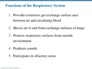
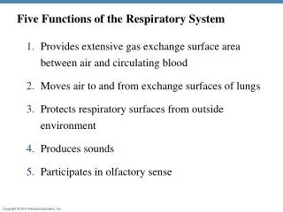
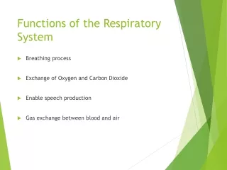
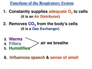
Functions of the Respiratory System. 3. Warms. air we breathe. 4. Filters. 5. Humidifies. 1. Constantly supplies adequate O 2 to cells. (it is an Air Distributor ). 2. Removes CO 2 from the body's cells. (it is a Gas Exchanger ). 6. Influences speech & sense of smell.

E N D
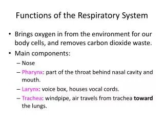
Functions of the Respiratory System 3. Warms air we breathe 4. Filters 5. Humidifies 1. Constantly supplies adequate O2 to cells (it is an Air Distributor) 2. Removes CO2 from the body's cells (it is a Gas Exchanger) 6. Influences speech & sense of smell
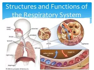
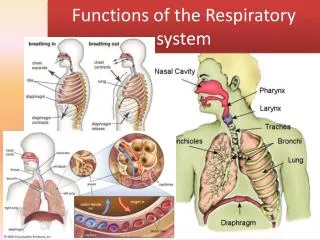
-- located outside the thoracic cavity & contains: nose pharynx larynx -- located in the thoracic cavity & contains: trachea bronchial tree lungs -- the Respiratory System has 2 divisions A. Upper Respiratory Tract (URT) B. Lower Respiratory Tract (LRT)
-- lines the air distribution tubes in the system -- covered with mucus & hair-like cilia -- this mucus is the -- traps foreign mostimportant air purification mechanism substances & moves them up & out Respiratory Mucosa
Respiratory Membrane capillary -- separates the air in the alveoli from the blood -- area where gas exchange occurs respiratory membrane alveoli
-- contains nerve endings for senseof smell Nose -- External nares (nostril) -- mainly a passageway for air -- warms & moistens inhaled air
Nose Ethmoid Frontal Sphenoid Maxillary -- mucosa that lines the sinuses is continuous with the mucosa that lines the nose Ethmoid -- also contains 4 paranasal sinuses: -- inflammation of the mucosa is called: Sinusitis
Pharynx Eustachian tube opening Nasopharynx Oropharynx Laryngopharynx -- opening to the Eustachian tubes are located in the Nasopharynx (allows for equalization of air pressures) -- why it is easy for a URI to turn into a middle ear infection -- Also called the Throat -- divided into 3 parts:
Epiglottis -- a tissue that covers the larynx while eating is the: Larynx -- composed of pieces of cartilage (largest piece called the Thyroid cartilageor "Adam's Apple")
Both Thyroid cartilage& vocal cords grow faster & larger in males at puberty -- 2 short fibrous bands that stretch across the interior of the Larynx Vocal Cords VOCAL CORDS -- if tensed, voice is high-pitched -- if relaxed, voice if low-pitched
-- is made of 15 - 20 C-shaped rings of cartilage Trachea -- these rings are almost non-collapsible -- also contains mucus glands TRACHEA -- extends from the larynx to the bronchi
-- even with the safeguard of non-collapsible rings, the trachea can be closed off due to: • aspiration of food or other objects -- complete obstruction can lead to death in minutes -- 5,000 people in the U.S. are killed each year TRACHEA a. tumors or infections
-- Right bronchus straighter than left, so aspirated foreign object tend to end up in right lung -- also contain rings of cartilage Bronchi -- contains 2 branches
-- do not contain rings of cartilage (made up of smooth muscle) Alveoli Bronchioles -- alveolar sacs are very thinned walled & lie close to thin walled blood capillaries (sacs that resemble grape clusters) -- this is where gas exchange occurs -- What 2 gases are involved here? O2 CO2
Venous Capillary Interstitial Space Alveoli Interstitial Space Lung Gas Exchange Rule: The Shorter the Distance through which Diffusion must take place, the Greater the Rate of Diffusion.
Alveoli Lung Gas Exchange Rule: The Greater the Surface Area which Diffusion Takes place, the Greater the Rate of Diffusion. --There are 300 Million Alveoli in the lungs surrounded by Capillaries.
-- the alveoli walls are lined with a lipoprotein called: Surfactant -- it reduces the surface tension in the alveoli -- reduces the lungs tendency to recoil Surfactant -- Premature infants may not have surfactant yet, so develop: Respiratory Distress Syndrome
Lungs -- Right lung contains 3 lobes Parietal Pleura Visceral Pleura Intrapleural space -- membrane that covers the lungs is called: -- area between the 2 membranes is called: -- Left lung only 2 Why? -- lining of the walls of the thoracic cavity is called: Inflammation of the Pleura is called: Pleurisy
Pneumothorax 761 760 -- pressure in the Intrapleural place should be less than atmospheric in order for lungs to remain inflated 754 Lung compressed Hole in lung Trachea Air in Pleural Space Chest wall Heart Diaphragm
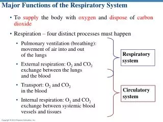
Breathing or Pulmonary Ventilation CO2 O2 CO2 O2 CO2 O2 O2 + food CO2 + ATP (exchange of gases between environment & alveoli) External Respirations (exchange of gases between the alveoli & the blood) Internal Respirations (exchange of gases between the blood & the cells) Cellular Respiration (using food & oxygen to make ATP)
Pulmonary ventilation -- because the lungs are enclosed in the Thoracic cavity,ifits shape is changed, the pressure within the cavity is changed -- the difference in air pressure between the atmosphere & the lungs is what causes air to move in or out of thelungs -- includes 2 phases: Inspiration (movement of air into the lungs) Expiration (movement of air out of the lungs)
-- changing the shape of the thoracic cavity involves 2 muscles: -- the most important muscle of inspiration 1. the Diaphragm 2. External Intercostals When the diaphragm contracts, it flattens out & makes the chest cavity longer from top to bottom -- the nerve that stimulates the diaphragm is the: Phrenic Nerve
-- the External Intercostals enlarges the thoracic cavity from front to back when contracted -- Expiration is primarily a passive action (These muscles just relax)
At Rest Pressure in Lungs Atmospheric Pressure Pressure in the Intrapleural Space INSPIRATION 760 Parietal Pleura Visceral Pleura AIR 754 760 760 754 Air moves from an area of higher pressure to an area of lower pressure 759
At Rest Pressure in Lungs Atmospheric Pressure Pressure in the Intrapleural Space 760 EXPIRATION 760 760 754 754 Air moves from an area of higher pressure to an area of lower pressure 761
-- During normal Expiration, the muscles of Inspiration relax -- However, when speaking, doing heavy work, --Decreases thoracic cavity from front to back -- push abdominal organs up against the diaphragm -- decreasing chest cavity from top to bottom -- this forces more air out past the vocal cords or singing, we need a more forceful expiration. -- the muscles involved here are the: 1. Internal Intercostals 2. Abdominal Muscles
Respiratory Control Centers are located in the: -- helps to maintain normal breathing rate of: Regulation of Breathing Medulla (with help from the Pons) 14 to 20 breaths/minute
-- can voluntarily speed up or slow down respirations -- we will resume normal breathing when body senses we need more O2 or CO2levels are too high -- children can hold their breath until they pass out (temper tantrum) -- however, will start to breathe normally once they do Cerebral Cortex
O2 CO2 -- thus increasing rate & depth of respirations Receptors that influence respiration 1. Chemoreceptors -- located: Carotid arteries Arch of the Aorta -- Stimulated by: Change in pH
-- located throughout the pulmonary airways & alveoli -- when the right amount of air enters the lungs, the receptors send a message to the Medulla to inhibit the Inspiratory Center 2. Pulmonary Stretch Receptors -- prevent over-inflation of the lungs
Liters 6 5 4 3 2.5 2 1 0 Time -- measures amounts of air exchanged in breathing Tidal Volume (TV) Tidal volume -- amount of air taken into & expelled in normal inspiration & expiration -- about 500 cc Spirometer
Liters 6 5 4 3 2.5 2 1 0 Time Inspiratory reserve (IRV) -- amount of air that can be forcibly inhaled over & above Tidal volume Inspiratory Tidal reserve volume -- about 3,000 cc
Liters 6 5 4 3 2.5 2 1 0 Time Expiratory reserve (ERV) Expiratory Inspiratory Tidal reserve reserve volume -- amount of air that can be forcibly exhaled over & above Tidal volume -- about 1000 cc
Liters 6 5 4 3 2.5 2 1 0 Time Vital Capacity (VC) -- largest amount of air that can be Expiratory Total Inspiratory Tidal lung reserve reserve Vital volume forcibly exhaled after maximum inhalation capacity capacity -- about 4500 cc (VC = IRV + ERV + TV)
Liters 6 5 4 3 2.5 2 1 Residual volume 0 Time Residual Volume Expiratory Total Inspiratory Tidal lung reserve reserve volume capacity -- air that remains in lungs after forcible exhalation -- about 1200 cc