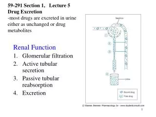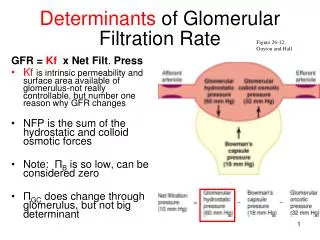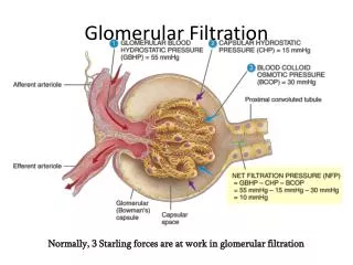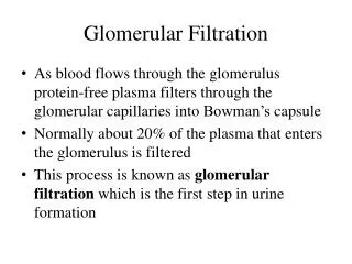
Glomerular Filtration
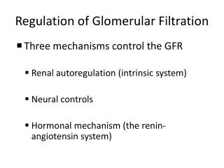
Glomerular Filtration. Control of the GFR Autoregulation (local level) Hormonal regulation (initiated by kidneys) Autonomic regulation (by sympathetic division of ANS). Glomerular Filtration. Autoregulation of the GFR Maintains GFR despite changes in local blood pressure and blood flow


Glomerular Filtration
E N D
Presentation Transcript
Glomerular Filtration • Control of the GFR • Autoregulation (local level) • Hormonal regulation (initiated by kidneys) • Autonomic regulation (by sympathetic division of ANS)
Glomerular Filtration • Autoregulation of the GFR • Maintains GFR despite changes in local blood pressure and blood flow • By changing diameters of afferent arterioles, efferent arterioles, and glomerular capillaries
Glomerular Filtration • Autoregulation of the GFR • Reduced blood flow or glomerular blood pressure triggers • Dilation of afferent arteriole • Dilation of glomerular capillaries • Constriction of efferent arterioles • Rise in renal blood pressure • Stretches walls of afferent arterioles • Causes smooth muscle cells to contract • Constricts afferent arterioles • Decreases glomerular blood flow
Glomerular Filtration • Hormonal Regulation of the GFR • By hormones of the • Renin–angiotensin system • Natriuretic peptides (ANP and BNP)
Glomerular Filtration • The Renin–Angiotensin System • Three stimuli cause the juxtaglomerular complex (JGA) to release renin • Decline in blood pressure at glomerulus due to decrease in blood volume • Fall in systemic pressures due to blockage in renal artery or tributaries • Stimulation of juxtaglomerular cells by sympathetic innervation due to decline in osmotic concentration of tubular fluid at macula densa
Glomerular Filtration • The Renin–Angiotensin System: Angiotensin II Activation • Constricts efferent arterioles of nephron • Elevating glomerular pressures and filtration rates • Stimulates reabsorption of sodium ions and water at PCT • Stimulates secretion of aldosterone by suprarenal (adrenal) cortex • Stimulates thirst • Triggers release of antidiuretic hormone (ADH) • Stimulates reabsorption of water in distal portion of DCT and collecting system
Glomerular Filtration • The Renin–Angiotensin System: Angiotensin II • Increases sympathetic motor tone • Mobilizing the venous reserve • Increasing cardiac output • Stimulating peripheral vasoconstriction • Causes brief, powerful vasoconstriction • Of arterioles and precapillary sphincters • Elevating arterial pressures throughout body
Glomerular Filtration • The Renin–Angiotensin System • Aldosterone • Accelerates sodium reabsorption: • in DCT and cortical portion of collecting system
Glomerular Filtration Figure 24–11a The Response to a Reduction in the GFR.
Glomerular Filtration Figure 24–11b The Response to a Reduction in the GFR.
Glomerular Filtration • Increased Blood Volume • Automatically increases GFR • To promote fluid loss • Hormonal factors further increase GFR • Accelerating fluid loss in urine
Glomerular Filtration • Natriuretic Peptides • Are released by the heart in response to stretching walls due to increased blood volume or pressure • Atrial natriuretic peptide (ANP) is released by atria • Brain natriuretic peptide (BNP) is released by ventricles • Trigger dilation of afferent arterioles and constriction of efferent arterioles • Elevates glomerular pressures and increases GFR
Glomerular Filtration • Autonomic Regulation of the GFR • Mostly consists of sympathetic postganglionic fibers • Sympathetic activation • Constricts afferent arterioles • Decreases GFR • Slows filtrate production • Changes in blood flow to kidneys due to sympathetic stimulation • May be opposed by autoregulation at local level
Reabsorption and Secretion • Reabsorption • Recovers useful materials from filtrate • Secretion • Ejects waste products, toxins, and other undesirable solutes • Both occur in every segment of nephron • Except renal corpuscle • Relative importance changes from segment to segment
Reabsorption and Secretion • Reabsorption and Secretion at the PCT • PCT cells normally reabsorb 60–70% of filtrate produced in renal corpuscle • Reabsorbed materials enter peritubular fluid • And diffuse into peritubular capillaries
Reabsorption and Secretion • Five Functions of the PCT • Reabsorption of organic nutrients • Active reabsorption of ions • Reabsorption of water • Passive reabsorption of ions • Secretion
Reabsorption and Secretion • Sodium Ion Reabsorption • Is important in every PCT process • Ions enter tubular cells by • Diffusion through leak channels • Sodium-linked cotransport of organic solutes • Countertransport for hydrogen ions
Reabsorption and Secretion Figure 24–12 Transport Activities at the PCT.
Reabsorption and Secretion • The Nephron Loop and Countercurrent Multiplication • Nephron loop reabsorbs about 1/2 of water and 2/3 of sodium and chloride ions remaining in tubular fluid by the process of countercurrent exchange
Reabsorption and Secretion • Countercurrent Multiplication • Is exchange that occurs between two parallel segments of loop of Henle • The thin, descending limb • The thick, ascending limb
Reabsorption and Secretion • Countercurrent • Refers to exchange between tubular fluids moving in opposite directions • Fluid in descending limb flows toward renal pelvis • Fluid in ascending limb flows toward cortex • Multiplication • Refers to effect of exchange • Increases as movement of fluid continues
Reabsorption and Secretion • Parallel Segments of Nephron Loop • Are very close together, separated only by peritubular fluid • Have very different permeability characteristics
Reabsorption and Secretion • The Thin Descending Limb • Is permeable to water • Is relatively impermeable to solutes • The Thick Ascending Limb • Is relatively impermeable to water and solutes • Contains active transport mechanisms • Pump Na+ and Cl- from tubular fluid into peritubular fluid of medulla
Reabsorption and Secretion • Sodium and Chloride Pumps • Elevate osmotic concentration in peritubular fluid • Around thin descending limb • Cause osmotic flow of water • Out of thin descending limb into peritubular fluid • Increasing solute concentration in thin descending limb
Reabsorption and Secretion • Concentrated Solution • Arrives in thick ascending limb • Accelerates Na+ and Cl- transport into peritubular fluid of medulla
Reabsorption and Secretion • Solute Pumping • At ascending limb • Increases solute concentration in descending limb • Which accelerates solute pumping in ascending limb
Reabsorption and Secretion • Countercurrent Multiplication • Active transport at apical surface • Moves Na+, K+ and Cl- out of tubular fluid • Uses carrier protein: Na+-K+/2 Cl- transporter
Reabsorption and Secretion • Na+-K+/2 Cl- Transporter • Each cycle of pump carries ions into tubular cell • 1 sodium ion • 1 potassium ion • 2 chloride ions
Reabsorption and Secretion Figure 24–13a Countercurrent Multiplication and Concentration of Urine.
Reabsorption and Secretion • Potassium Ions • Are pumped into peritubular fluid by cotransport carriers • Are removed from peritubular fluid by sodium–potassium exchange pump • Diffuse back into lumen of tubule through potassium leak channels
Reabsorption and Secretion • Sodium and Chloride Ions • Removed from tubular fluid in ascending limb • Elevate osmotic concentration of peritubular fluid around thin descending limb
Reabsorption and Secretion • The Thin Descending Limb • Is permeable to water, impermeable to solutes • As tubular fluid flows along thin descending limb • Osmosis moves water into peritubular fluid, leaving solutes behind • Osmotic concentration of tubular fluid increases
Reabsorption and Secretion Figure 24–13b Countercurrent Multiplication and Concentration of Urine.
Reabsorption and Secretion • The Thick Ascending Limb • Has highly effective pumping mechanism • 2/3 of Na+ and Cl- are pumped out of tubular fluid before it reaches DCT • solute concentration in tubular fluid declines
Reabsorption and Secretion Figure 24–13c Countercurrent Multiplication and Concentration of Urine.
Reabsorption and Secretion • Tubular Fluid at DCT • Arrives with osmotic concentration of 100 mOsm/L • 1/3 concentration of peritubular fluid of renal cortex • Rate of ion transport across thick ascending limb is proportional to ion’s concentration in tubular fluid
Reabsorption and Secretion • Regional Differences • More Na+ and Cl- are pumped into medulla • At start of thick ascending limb than near cortex • Regional difference in ion transport rate • Causes concentration gradient within medulla
Reabsorption and Secretion • The Concentration Gradient of the Medulla • Of peritubular fluid near turn of nephron loop • 1200 mOsm/L: • 2/3 (750 mOsm/L) from Na+ and Cl-: • pumped out of ascending limb • remainder from urea
Reabsorption and Secretion • Urea and the Concentration Gradient • Thick ascending limb of nephron loop, DCT, and collecting ducts are impermeable to urea • As water is reabsorbed, concentration of urea rises • Tubular fluid reaching papillary duct contains 450 mOsm/L urea • Papillary ducts are permeable to urea • Concentration in medulla averages 450 mOsm/L
Reabsorption and Secretion • Benefits of Countercurrent Multiplication • Efficiently reabsorbs solutes and water: • Before tubular fluid reaches DCT and collecting system • Establishes concentration gradient: • That permits passive reabsorption of water from tubular fluid in collecting system: • regulated by circulating levels of antidiuretic hormone (ADH)
Reabsorption and Secretion • Reabsorption and Secretion at the DCT • Composition and volume of tubular fluid • Changes from capsular space to distal convoluted tubule: • only 15–20% of initial filtrate volume reaches DCT • concentrations of electrolytes and organic wastes in arriving tubular fluid no longer resemble blood plasma
Reabsorption and Secretion • Reabsorption at the DCT • Selective reabsorption or secretion, primarily along DCT, makes final adjustments in solute composition and volume of tubular fluid • Tubular Cells at the DCT • Actively transport Na+ and Cl- out of tubular fluid • Along distal portions: • contain ion pumps • reabsorb tubular Na+ in exchange for K+
Reabsorption and Secretion • Aldosterone • Is a hormone produced by suprarenal cortex • Controls ion pump and channels • Stimulates synthesis and incorporation of Na+ pumps and channels • In plasma membranes along DCT and collecting duct • Reduces Na+ lost in urine
Reabsorption and Secretion • Hypokalemia • Produced by prolonged aldosterone stimulation • Dangerously reduces plasma concentration
Reabsorption and Secretion • Natriuretic Peptides (ANP and BNP) • Oppose secretion of aldosterone • And its actions on DCT and collecting system • Parathyroid Hormone and Calcitriol • Circulating levels regulate reabsorption at the DCT
Reabsorption and Secretion • Secretion at the DCT • Blood entering peritubular capillaries • Contains undesirable substances that did not cross filtration membrane at glomerulus • Rate of K+ and H+ secretion rises or falls • According to concentrations in peritubular fluid • Higher concentration and higher rate of secretion
Reabsorption and Secretion • Potassium Ion Secretion • Ions diffuse into lumen through potassium channels • At apical surfaces of tubular cells • Tubular cells exchange Na+ in tubular fluid • For excess K+ in body fluids
Reabsorption and Secretion Figure 24–14a, b Tubular Secretion and Solute Reabsorption at the DCT.
Reabsorption and Secretion Figure 24–14a, b Tubular Secretion and Solute Reabsorption at the DCT.