22
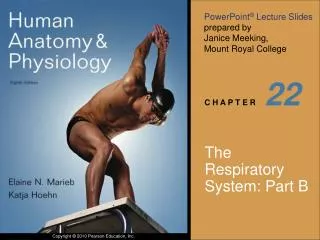
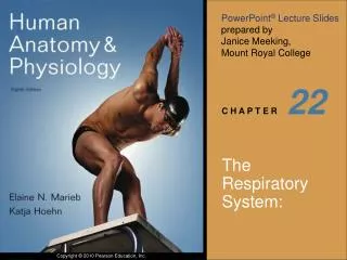
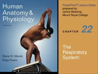
22 . The Respiratory System: Part A. Nasal cavity. Oral cavity. Nostril. Pharynx. Larynx. Left main (primary) bronchus. Trachea. Carina of trachea. Right main (primary) bronchus. Left lung. Diaphragm. Right lung. Figure 22.1. Functional Anatomy.

22
E N D
Presentation Transcript
22 The Respiratory System:Part A
Nasal cavity Oral cavity Nostril Pharynx Larynx Left main (primary) bronchus Trachea Carina of trachea Right main (primary) bronchus Left lung Diaphragm Right lung Figure 22.1
Functional Anatomy • Respiratory zone: site of gas exchange • Microscopic structures: respiratory bronchioles, alveolar ducts, and alveoli • Conducting zone: conduits to gas exchange sites • Includes all other respiratory structures • Respiratory muscles: diaphragm and other muscles that promote ventilation
Respiratory mucosa • Pseudostratified ciliated columnar epithelium • Mucous and serous secretions contain lysozyme and defensins • Cilia move contaminated mucus posteriorly to throat • Inspired air is warmed by plexuses of capillaries and veins
Mucosa • Pseudostratified ciliated columnar epithelium • Lamina propria (connective tissue) Submucosa Seromucous gland in submucosa Hyaline cartilage (b) Photomicrograph of the tracheal wall (320x) Figure 22.6b
Pharynx Nasopharynx Oropharynx Laryngopharynx (b) Regions of the pharynx Figure 22.3b
Larynx • Attaches to the hyoid bone and opens into the laryngopharynx • Continuous with the trachea • Functions • Provides a patent airway • Routes air and food into proper channels • Voice production
Larynx • Vocal ligaments • Attach the arytenoid cartilages to the thyroid cartilage • Contain elastic fibers • Form core of vocal folds (true vocal cords) • Opening between them is the glottis • Folds vibrate to produce sound as air rushes up from the lungs
Larynx • Vestibular folds (false vocal cords) • Superior to the vocal folds • No part in sound production • Help to close the glottis during swallowing
Base of tongue Epiglottis Vestibular fold (false vocal cord) Vocal fold (true vocal cord) Glottis Inner lining of trachea Cuneiform cartilage Corniculate cartilage (a) Vocal folds in closed position; closed glottis (b) Vocal folds in open position; open glottis Figure 22.5
Conducting Zone Structures • Trachea right and left main (primary) bronchi • Each main bronchus enters the hilum of one lung • Right main bronchus is wider, shorter, and more vertical than the left • Each main bronchus branches into lobar (secondary) bronchi (three right, two left) • Each lobar bronchus supplies one lobe
Conducting Zone Structures • Each lobar bronchus branches into segmental (tertiary) bronchi • Segmental bronchi divide repeatedly • Bronchioles are less than 1 mm in diameter • Terminal bronchioles are the smallest, less than 0.5 mm diameter
Conducting Zone Structures • From bronchi through bronchioles, structural changes occur • Cartilage rings give way to plates; cartilage is absent from bronchioles • Epithelium changes from pseudostratified columnar to cuboidal; cilia and goblet cells become sparse • Relative amount of smooth muscle increases
Respiratory Zone • Respiratory bronchioles, alveolar ducts, alveolar sacs (clusters of alveoli) • ~300 million alveoli account for most of the lungs’ volume and are the main site for gas exchange
Alveoli Alveolar duct Respiratory bronchioles Alveolar duct Terminal bronchiole Alveolar sac (a) Figure 22.8a
Respiratory Membrane • ~0.5-m-thick air-blood barrier • Alveolar and capillary walls and their fused basement membranes • Alveolar walls • Single layer of squamous epithelium (type I cells) • Scattered type II cuboidal cells secrete surfactant and antimicrobial proteins
Alveoli • Surrounded by fine elastic fibers • Contain open pores that • Connect adjacent alveoli • Allow air pressure throughout the lung to be equalized • House alveolar macrophages that keep alveolar surfaces sterile
Red blood cell Nucleus of type I (squamous epithelial) cell Alveolar pores Capillary O2 Capillary Type I cell of alveolar wall CO2 Alveolus Macrophage Alveolus Endothelial cell nucleus Alveolar epithelium Fused basement membranes of the alveolar epithelium and the capillary endothelium Respiratory membrane Red blood cell in capillary Alveoli (gas-filled air spaces) Type II (surfactant- secreting) cell Capillary endothelium (c) Detailed anatomy of the respiratory membrane Figure 22.9c
Blood Supply • Pulmonary circulation (low pressure, high volume) • Pulmonary arteries deliver systemic venous blood • Branch profusely, along with bronchi • Feed into the pulmonary capillary networks • Pulmonary veins carry oxygenated blood from respiratory zones to the heart
Blood Supply • Systemic circulation (high pressure, low volume) • Bronchial arteries provide oxygenated blood to lung tissue • Arise from aorta and enter the lungs at the hilum • Supply all lung tissue except the alveoli • Bronchial veins anastomose with pulmonary veins • Pulmonary veins carry most venous blood back to the heart
Pleurae • Thin, double-layered serosa • Parietal pleura on thoracic wall and superior face of diaphragm • Visceral pleura on external lung surface • Pleural fluid fills the slitlike pleural cavity • Provides lubrication and surface tension
Mechanics of Breathing • Pulmonary ventilation consists of two phases • Inspiration: gases flow into the lungs • Expiration: gases exit the lungs
Atmospheric pressure Parietal pleura Thoracic wall Visceral pleura Pleural cavity Transpulmonary pressure 760 mm Hg –756 mm Hg = 4 mm Hg 756 Intrapleural pressure 756 mm Hg (–4 mm Hg) 760 Intrapulmonary pressure 760 mm Hg (0 mm Hg) Lung Diaphragm Figure 22.12
Pulmonary Ventilation • Inspiration and expiration • Mechanical processes that depend on volume changes in the thoracic cavity • Volume changes pressure changes • Pressure changes gases flow to equalize pressure
Boyle’s Law • The relationship between the pressure and volume of a gas • Pressure (P) varies inversely with volume (V): P1V1 = P2V2
Changes in lateral dimensions (superior view) Changes in anterior- posterior and superior- inferior dimensions Sequence of events 1 Inspiratory muscles contract (diaphragm descends; rib cage rises). Ribs are elevated and sternum flares as external intercostals contract. Thoracic cavity volume increases. 2 External intercostals contract. 3 Lungs are stretched; intrapulmonary volume increases. Intrapulmonary pressure drops (to –1 mm Hg). 4 5 Air (gases) flows into lungs down its pressure gradient until intrapulmonary pressure is 0 (equal to atmospheric pressure). Diaphragm moves inferiorly during contraction. Figure 22.13 (1 of 2)
Changes in lateral dimensions (superior view) Changes in anterior- posterior and superior- inferior dimensions Sequence of events 1 Inspiratory muscles relax (diaphragm rises; rib cage descends due to recoil of costal cartilages). Ribs and sternum are depressed as external intercostals relax. 2 Thoracic cavity volume decreases. 3 Elastic lungs recoil passively; intrapulmonary volume decreases. External intercostals relax. 4 Intrapulmonary pres- sure rises (to +1 mm Hg). Diaphragm moves superiorly as it relaxes. 5 Air (gases) flows out of lungs down its pressure gradient until intra- pulmonary pressure is 0. Figure 22.13 (2 of 2)
Intrapulmonary pressure. Pressure inside lung decreases as lung volume increases during inspiration; pressure increases during expiration. Inspiration Expiration Intrapulmonary pressure Trans- pulmonary pressure Intrapleural pressure. Pleural cavity pressure becomes more negative as chest wall expands during inspiration. Returns to initial value as chest wall recoils. Intrapleural pressure Volume of breath Volume of breath. During each breath, the pressure gradients move 0.5 liter of air into and out of the lungs. 5 seconds elapsed Figure 22.14
Physical Factors Influencing Pulmonary Ventilation • Inspiratory muscles consume energy to overcome three factors that hinder air passage and pulmonary ventilation • Airway resistance • Alveolar surface tension • Lung compliance
Conducting zone Respiratory zone Medium-sized bronchi Terminal bronchioles Airway generation (stage of branching) Figure 22.15
Alveolar Surface Tension • Surface tension • Attracts liquid molecules to one another at a gas-liquid interface • Resists any force that tends to increase the surface area of the liquid
Alveolar Surface Tension • Surfactant • Detergent-like lipid and protein complex produced by type II alveolar cells • Reduces surface tension of alveolar fluid and discourages alveolar collapse • Insufficient quantity in premature infants causes infant respiratory distress syndrome
Lung Compliance • A measure of the change in lung volume that occurs with a given change in transpulmonary pressure • Normally high due to • Distensibility of the lung tissue • Alveolar surface tension
Lung Compliance • Diminished by • Nonelastic scar tissue (fibrosis) • Reduced production of surfactant • Decreased flexibility of the thoracic cage
22 The Respiratory System: Part B
Respiratory Volumes • Used to assess a person’s respiratory status • Tidal volume (TV) • Inspiratory reserve volume (IRV) • Expiratory reserve volume (ERV) • Residual volume (RV)
Adult female average value Adult male average value Measurement Description Amount of air inhaled or exhaled with each breath under resting conditions Tidal volume (TV) 500 ml 500 ml Amount of air that can be forcefully inhaled after a nor- mal tidal volume inhalation Inspiratory reserve volume (IRV) 3100 ml 1900 ml Respiratory volumes Amount of air that can be forcefully exhaled after a nor- mal tidal volume exhalation Expiratory reserve volume (ERV) 1200 ml 700 ml Amount of air remaining in the lungs after a forced exhalation Residual volume (RV) 1200 ml 1100 ml Figure 22.16b
Respiratory Capacities • Inspiratory capacity (IC) • Functional residual capacity (FRC) • Vital capacity (VC) • Total lung capacity (TLC)
Maximum amount of air contained in lungs after a maximum inspiratory effort: TLC = TV + IRV + ERV + RV Total lung capacity (TLC) 6000 ml 4200 ml Maximum amount of air that can be expired after a maxi- mum inspiratory effort: VC = TV + IRV + ERV Vital capacity (VC) 4800 ml 3100 ml Respiratory capacities Maximum amount of air that can be inspired after a normal expiration: IC = TV + IRV Inspiratory capacity (IC) 3600 ml 2400 ml Volume of air remaining in the lungs after a normal tidal volume expiration: FRC = ERV + RV Functional residual capacity (FRC) 2400 ml 1800 ml (b) Summary of respiratory volumes and capacities for males and females Figure 22.16b
Inspiratory reserve volume 3100 ml Inspiratory capacity 3600 ml Vital capacity 4800 ml Total lung capacity 6000 ml Tidal volume 500 ml Expiratory reserve volume 1200 ml Functional residual capacity 2400 ml Residual volume 1200 ml (a) Spirographic record for a male Figure 22.16a
Dead Space • Some inspired air never contributes to gas exchange • Anatomical dead space: volume of the conducting zone conduits (~150 ml) • Alveolar dead space: alveoli that cease to act in gas exchange due to collapse or obstruction • Total dead space: sum of above nonuseful volumes
Pulmonary Function Tests • Increases in TLC, FRC, and RV may occur as a result of obstructive disease • Reduction in VC, TLC, FRC, and RV result from restrictive disease
Basic Properties of Gases • The amount of gas that will dissolve in a liquid also depends upon its solubility • CO2 is 20 times more soluble in water than O2 • Very little N2 dissolves in water
Inspired air: P 160 mm Hg P 0.3 mm Hg Alveoli of lungs: P 104 mm Hg P 40 mm Hg O2 O2 CO2 CO2 External respiration Pulmonary arteries Pulmonary veins (P 100 mm Hg) O2 Blood leaving tissues and entering lungs: P 40 mm Hg P 45 mm Hg Blood leaving lungs and entering tissue capillaries: P 100 mm Hg P 40 mm Hg O2 O2 CO2 CO2 Heart Systemic veins Systemic arteries Internal respiration Tissues: P less than 40 mm Hg P greater than 45 mm Hg O2 CO2 Figure 22.17
O2 Transport • Molecular O2 is carried in the blood • 1.5% dissolved in plasma • 98.5% loosely bound to each Fe of hemoglobin (Hb) in RBCs • 4 O2 per Hb
O2 and Hemoglobin • Rate of loading and unloading of O2 is regulated by • Po2 • Temperature • Blood pH • Pco2 • Concentration of BPG
Influence of Po2 on Hemoglobin Saturation • Oxygen-hemoglobin dissociation curve • Hemoglobin saturation plotted against Po2 is not linear • S-shaped curve • Shows how binding and release of O2 is influenced by the Po2
O2 unloaded to resting tissues Additional O2 unloaded to exercising tissues Exercising tissues Resting tissues Lungs Figure 22.20
Other Factors Influencing Hemoglobin Saturation • Increases in temperature, H+, Pco2, and BPG • Modify the structure of hemoglobin and decrease its affinity for O2 • Occur in systemic capillaries • Enhance O2 unloading • Shift the O2-hemoglobin dissociation curve to the right • Decreases in these factors shift the curve to the left
Decreased carbon dioxide (P 20 mm Hg) or H+ (pH 7.6) 10°C CO2 20°C 38°C 43°C Normal arterial carbon dioxide (P 40 mm Hg) or H+ (pH 7.4) CO2 Normal body temperature Increased carbon dioxide (P 80 mm Hg) or H+ (pH 7.2) CO2 (a) P (mm Hg) O2 (b) Figure 22.21