CROHN DESEASE New Imaging
520 likes | 699 Vues
CROHN DESEASE New Imaging. BEN ROMDHANE MH Hopital AVICENNE BOBIGNY. role cross-sectional imaging expanded CT and MRI allow rapid acquisition of high-resolution images of the intestines. Protocoles necessary to acquire images of diagnostic quality

CROHN DESEASE New Imaging
E N D
Presentation Transcript
CROHN DESEASENew Imaging BEN ROMDHANE MH Hopital AVICENNE BOBIGNY
role cross-sectional imaging expanded • CT and MRI allow rapid acquisition of high-resolution images of the intestines. • Protocoles necessary to acquire images of diagnostic quality • Sensitivity (CT and MR I ) of over 95% for the detection of CD
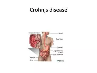
Chronic granulomatous GI tract inflammation • peakbetween 15 and 25 years of age • tendency toward remission and relapse • affect any part of the GI tract • often multiple discontinuous sites • Small intestine 80% of cases, • terminal ileum most commonly • Colon affected with (50% of cases) or without (15%–20%) involvement of the small intestine
Earliestchange in the submucosa with lymphoid hyperplasia and lymphedema • Early stage: subtle elevations and aphthoid ulcers. • Transmurally extension to serosa (transmural stage) and • Extension to mesentery and adjacent organs (extramural stage). • Aphthoid ulcers develop into linear ulcers and fissures to produce an ulceronodularor “cobblestone” appearance.
bowelwall thickened by fibrosis and / or inflammatory infiltrates • common complications of advanced disease: Bowel obstruction, strictures, abscesses or phlegmon, fistulas, and sinus tracts • not common, toxic megacolon and neoplasms (lymphoma and carcinoma)
Endoscopy and barium studies limited in demonstrating the transmural or extramural extent or extraintestinal complications • Cross-sectional imaging may not detect subtle mucosal lesions • but reveals pathologic changes of the intestinal mucosa • help compensate for the limitations of conventional imaging
CT currently the cross-sectional imaging modality of choice at most institutions • MRI has also proved highly effective • CT and MRI allow rapid acquisition of high-resolution images of the intestines during a breath-hold examination. • CT and MRI provide useful information in the diagnosis and in treatment planning
limited spatial resolution of CT and MRI compared with enteroclysis studies results in lower rates of depiction of early disease manifestations • Detection and characterization of intestinal lesionsrequireappropriatepreparation and scanning techniques. • GI tract should be empty and clean, with the lumen distended
review preparations, contrast agents, and scanning techniques • illustrate the characteristic imaging appearances of CD • Review findings that indicate the presence of inflammatory lesion activity • discuss advantages, limitations and role of cross-sectional imaging
Fast for 6 H priorexamination (decreases the alimentaryresidue • Collapsedbowel loops may mimic a segment with wall thickening, an abscess, or enlarged lymph nodes • Administration of large amount of intraluminal contrast distends bowel loops for better visualisation of anatomie and morphologic changes • IV contrast demonstrate the presence of lesions and help assess their inflammatory activity
Advantages of cross-sectional imaging • demonstrate : transmural extent of inflammation, skip lesions beyond severe luminal stenoses, intraperitoneal or extraintestinal complications • provide three-dimensional information • and, vascular information with use of contrast material
Entéroscanner Ingestion soluté hyperosmolaire/ Sonde entéroclyse sous scopie dans D3 Antispasmodique IV Scanner sans et après injection IV produit de contraste
The contrast agent should allow imaging with - homogeneous luminal enhancement -high contrast between lumen and bowel wall -minimal mucosal absorption - absence of artifact formation - no significant adverse effects
To minimize bowel movement or contraction and motion artifact from intestinal peristalsis: - Antiperistaltic agents prior to scanning - short scanning time • Intravenous contrast medium indicated for : - better visualization of the bowel wall extraintestinal structures, and lesions - precise evaluation of the degree of inflammatory activity
ComputedTomography • Various types of intraluminal contrast media are used to provide positive or negative contrast between the bowel lumen and surrounding structures • Positive contrast agent with high attenuation at CT aids in differentiating bowel loops from enlarged lymph nodes or an extramural fluid collection such as an abscess. • The presence of small bowel obstruction or fistulas is also well appreciated.
with the use of positive intraluminal contrast material, mural enhancementafter iv injection may obscure subtle Crohn lesions. • The use of negative intraluminal contrast agents with low attenuation facilitates depiction of the wall of normal and diseased bowel segments, particularly after iv contrastmaterial administration
technique for intraluminal contrast material administration is common to both CT and MRI • Between 1,500 and 2,000 mL of contrast material administered orally 45–90 minutes prior to the examination • To provide adequate and uniform distention of the bowel loops, patients are asked to steadily ingest the contrast material over a 20–60-minutes period
The contrastmaterialmay be administered through a nasojejunal catheter at a rate of 100–250 mL/min with thehelp of a roller pump • CT or MR imaging performed with this technique is called CT or MR enteroclysis • Use of a nasojejunal catheter allows better luminal distention but causes patient discomfort • If necessary, 300– 1,000 mL of contrast agent can be administered transrectal
CT scans with the patient in the prone position is recommended to disperse the small bowel loops • With multi–detector row CT scanners, thinner collimation (0.5–2.5 mm) is possible. Sections with a 5–7-mm thickness or thinner sections, overlapping reconstructed images, or multiplanar reformatted images • IV administration of iodinated contrast essential, 100–150 ml at a rate of 2.5–4ml /sec with a delay time of 40–70 sec
MR Imaging • Various kinds of intraluminal contrast agents positive, negative, or biphasic • Positive agents produce high intraluminal signal • Negative agents produce little or no intraluminal signal • Biphasic contrast agents may produce either high or low signal depending on the pulse sequence used, usually demonstrating low signal intensity on T1 and high signal on T2 • Negative or biphasic agents more suitable
An antiperistaltic agent injected to minimize potential artifact of bowel movement or contraction • Prone position recommended for separating bowel loops and decreasing the scanning volume. This position is also safe for patients should they vomit
To increase the signal-to-noise ratio, use of abdominal phased array radiofrequency coils • Coronal images obtained with a 4–7-mm section thickness, a 128–256 256 matrix, and a field of view of 350 mm or more • Thicker sections to monitor the infusion process ( 70–180 mm) • Acquisition of axial, sagittal, or multiplanar images may be necessary for precise evaluation
Protocol should include both T1- and T2- to detect and characterize each lesion • T1-weighted imaging with iv contrast essential for assessing inflammatory lesion activity. • True fast imaging with a steady precession (FISP), • half-Fourier acquired single shot fast spin-echo, • T2-weighted turbo spin-echo, • combination of these sequences recommended
Gadolinium enhanced fat-suppressedspoiled gradient-echo T1 2D ou 3D • excellent visualization of the enhancing bowel wall ( contrasts with the low signal mesenteric fat and negativintraluminalcontrastmaterial) • Morphologicfeatures and degree of enhancement both aid in assessing CD activity • Images covering the bowel loops in their entirety can be obtained within 30 sec • Scanning is performed after a bolus iv injection of 0.1–0.2 mmol/kg of gadopentetatedimeglumine with a delay time of 40–80 sec
CT and MR Imaging Findings in CD In proved or suspected CD images analyzed specifically for : - altered bowel segment(wall thickness, attenuation , degree of enhancement, length of involvement) - stenosis and prestenotic dilatation - skip lesions, fistulas, abscess, fibrofattyproliferation, increased vascularity of the vasa recta (comb sign), mesenteric adenopathy, and other extra-intestinal disease involvement
Normal thickness of the wall of the small intestine1–2 mm and colon 3 mm when lumen is distended • Any portion of the bowel wall that exceeds 4–5 mm is considered abnormal • Bowel wall thickening, usually ranging from 1–2 cm, is the most consistent feature of CD
number of lesions and extent of Involvement • involved segment homogeneous or stratified appearance (alternating layers of higher or lower attenuation or signal intensity) CT / MRI
Mural stratification (“target” or “double halo” appearance) often seen in active lesions after iv contrast • inflamedbowelwalldemonstratesmarkedenhancementafter iv contrast • intensity of enhancement correlates with degree of inflammatory lesion activity
normal small intestine lumen less than 2.5 cm • Luminal narrowing and associated prestenotic dilatation easily recognized • Deformity of bowel loops such as pseudo-diverticulum formation caused by asymmetric involvement by longitudinal ulcers and ulcer scars is well demonstrated on both axial and coronal images.
early-stage lesions such as enlarged lymph follicles, slight distortion of the bowel folds, and tiny aphtae are not consistently visible at either CT or MRI due to inadequate spatial resolution
Fibrofatty proliferation of the mesentery is commonly seen adjacent to involved bowel segment in CD • CT and MRI demonstrate fibrofatty proliferation, • which has slightly increased CT attenuation and • slightly decreased MRI signal intensity compared with normal fat separating the bowel loops.
Abscess and phlegmon well demonstrated at CT and fat-saturated T2-MRI • can occur in small bowel mesentery abdominal wall psoas muscle or around the anus • Fistulas and sinus tracts are also depicted • MRI sensitivity for depicting sinus tracts is 50%–75% /conventional enteroclysis
Mesenteric lymphadenopathy ranging from 3 to 8 mm in size depicted at CT and MRI • When lymph nodes larger than 10 mm, lymphoma and carcinoma must beexcluded.
Inflammatory activity • well appreciated at CT and MR imaging • Findings include : thickened bowel wall with marked contrast enhancement, mural stratification, pericolicor perienterichypervascularity (comb sign) hyperintensity T 2 of the bowel wall lymphnodeenlargement, extramural complications: phlegmon abscess
CT sensitivity 94%–100% specificity 95% • Sensitivity increases to 98% in the diagnosis of transmural or extramural • only 70% for early-stage disease. • multiplanar images with axial images significantly improves observer confidence • Sensitivity of MRI 96%–100% • sensitivity on a per lesion 85 % and 100% when superficial lesions excluded