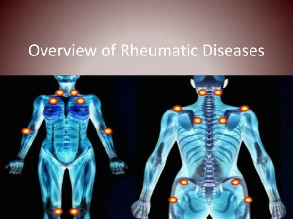
INTRODUCTION TO RHEUMATOLOGY
970 likes | 3.61k Vues
INTRODUCTION TO RHEUMATOLOGY. KATHRYN DAO, MD Arthritis Consultation Center July 21, 2005. What am I? I am the size of a rabbit with fur as smooth as an otter. I have a spongy beak covered with sensitive skin. I am a monotreme.

INTRODUCTION TO RHEUMATOLOGY
E N D
Presentation Transcript
INTRODUCTION TO RHEUMATOLOGY KATHRYN DAO, MD Arthritis Consultation Center July 21, 2005
What am I? I am the size of a rabbit with fur as smooth as an otter. I have a spongy beak covered with sensitive skin. I am a monotreme. I protect myself with poisonous spurs from my hind legs My name means "flat feet."
Why even care? 3500 rheumatologists • 2002 CDC reported arthritis as the leading cause of disability in the US. • 55.4 million have chronic joint symptoms lasting for more than 3 months • 21.5 million have not seen a physician • 2 million have activity limitations • 25% will be unable to work within 7 years of disease onset • Direct and indirect costs are estimated at 1% of the US gross domestic product = $86.2 billion Center for Disease Control and Prevention. MMWR 2004;53:383-6. Center for Disease Control and Prevention. MMWR 2004;53:388.
Musculoskeletal Complaint Joint Pain Joint Swelling Diffuse/Systemic Sxs • Initial Rheumatic History and Physical Exam to Determine: • 1. Is it articular • 2. Is it acute or chronic? • 3. Is inflammation present? • 4. How many/which joints are involved? • 5. Are there RED FLAGS?
Goals of Assessment • Identify “Red Flag” conditions • Conditions with sufficient morbidity/mortality to warrant an expedited diagnosis • Make a timely diagnosis • Common conditions occur commonly • Many SkM conditions are self-limiting • Some conditions require serial evaluation over time to make a Dx • Provide relief, reassurance and plan for evaluation and treatment
RED FLAG CONDITIONS • FRACTURE • INFECTION • ORGAN INVOLVEMENT
Articular vs. Periarticular Finding ARTICULAR PERIARTICULAR PainDiffuse, deep "point" tenderness ROM PainActive+passive Active motion in all planes in few planes Swelling Common Uncommon
Peri-/Non-articular Pain • Fibromyalgia • Fracture • Bursitis, Tendinitis, Enthesitis, Periostitis • Carpal tunnel syndrome • Polymyalgia rheumatica • Sickle Cell Crisis • Raynaud’s phenomenon • Reflex sympathetic dystrophy • Myxedema
Less than 4 joints Osteoarthritis Fracture Osteonecrosis Gout or Pseudogout Septic arthritis Lyme disease Reactive arthrtis Tuberculous/Fungal arthritis Sarcoidosis 4 or more joints Osteoarthritis Rheumatoid arthritis Psoriatic arthritis Viral arthritis Serum Sickness Juvenile arthritis SLE/PSS/MCTD Mono/Oligo vs Polyarticular
History: Clues to Diagnosis • Age • Young: JRA, SLE, Reiter's, GC arthritis • Middle: Fibromyalgia, tendinitis, bursitis, LBP RA • Elderly: OA, crystals, PMR, septic, osteoporosis • Sex • Males: Gout, AS, Reiter's syndrome • Females: Fibrositis, RA, SLE, osteoarthritis • Race • White: PMR, GCA and Wegener's • Black: SLE, sarcoidosis • Asian: RA, SLE, Takayasu's arteritis, Behcet's
Rheumatic Review of Systems • Constitutional: fever, wt loss, fatigue • Ocular: blurred vision, diplopia, conjunctivitis, dry eyes • Oral: dental caries, ulcers, dysphagia, dry mouth • GI: hx ulcers, Abd pain, change in BM, melena, jaundice • Pulm: SOB, DOE, hemoptysis, wheezing • CVS: angina/CP, arrhythmia, HTN, Raynauds • Skin: photosensitivity, alopecia, nails, rash • CNS: HA, Sz, weakness, paraesthesias • Reproductive: sexual dysfunction, promiscuity, genital lesions, miscarriages, impotence • SkM: joint pain/swelling, stiffness, ROM/function, nodules
Rheumatic Review of Systems • Fever/Constitutional: septic arthritis, vasculitis, Still’s disease • Ocular: Reiters, Behcets, Sjogrens, Cataracts (steroids) • Oral: Sjogrens, Lupus, GC, myositis, drugs • GI: Reactive arthritis, IBD, hepatitis, Polyarteritis, Scleroderma • Pulm: SLE, RA lung, Churg-Strauss, Wegeners, Scleroderma • CVS: Vasculitis, PSS, Raynauds, antiphospholipid syndrome • Skin: SLE, psoriatic, vasculitis, Kawasaki syndrome • CNS: lupus carpal tunnel, antiphospholipid, vasculitis • GYN/GU: antiphospholipid, SLE, Reiters, Behcets, CTX • Musculoskeletal: Gout, RA, OA, fibromyalgia, fracture
Onset & Chronology • Acute: Fracture, septic arthritis, gout, rheumatic fever, Reiter's syndrome • Chronic: OA, RA, SLE, psoriatic arthritis, fibromyalgia • Intermittent: gout, pseudogout, Lyme, palindromic rheumatism, Behcet's, Familial Mediterranean Fever • Additive: OA, RA, Reiter's syndrome, psoriatic • Migratory: Viral arthritis (hepatitis B), rheumatic fever, GC arthritis, SLE
Initial Rheumatic History and Physical Exam to Determine: • 1. Is it articular • 2. Is it acute or chronic? • 3. Is inflammation present? • 4. How many/which joints are involved? Musculoskeletal Complaint • Nonarticular Condition • Trauma/Fracture • Fibromyalgia • Polymyalgia Rheumatica • Bursitis • Tendinitis Is it Articular? No Yes Is Complaint > 6 wks Duration? Yes No Is Inflammation Present? 1. Is there prolonged AM stiffness? 2. Is there soft tissue swelling? 3. Are there systemic symptoms? 4. Is the ESR or CRP elevated? • Acute Arthritis • Infectious Arthritis • Gout • Pseudogout • Reiter’s Syndrome • Initial Presentation of Chronic Arthritis Chronic Acute No Yes Chronic Inflammatory Arthritis • Chronic Inflammatory • Mono/oligoarthritis • Consider: • Indolent infection • Psoriatic Arthritis • Reiter’s Syndrome • Pauciarticular JA Chronic Noninflammatory Arthritis <4 How Many Joints Involved? Are DIP, CMC, Hip or Knee Involved? 4+ Chronic Inflammatory Polyarthritis No Yes No • Consider: • Psoriatic Arthritis • Reiter’s Syndrome Is it Symmetric? • Unlikely to be • Osteoarthritis • Consider: • Osteonecrosis • Charcot Arthritis Osteoarthritis Yes • Consider: • SLE • Scleroderma • Polymyositis Rheumatoid Arthritis Are PIP, MCP or MTP Joints Involved? No Yes Adapted from J. Cush, MD
Know It When You See It • Hard bony enlargements • Heberden’s nodes at the DIP joints • Bouchard’s nodes at the PIP joints • Often have “squared” first CMC joint due to osteophytes at that joint Osteoarthritis
Know It When You See It • Soft synovial swelling • Synovitis and volar subluxation at the MCP joints • Synovitis of the wrists • Synovitis of the PIP joints with early swan neck deformities Rheumatoid arthritis
Rheumatoid Arthritis: Late Stages • Deformities • Nodules • Tendon Rupture
Know It When You See It Jaccoud’s Deformity of SLE
Know It When You See It • Often associated with: • Inflammatory eye disease • Balanitis, oral ulceration, or keratoderma • Enthesopathy • Sacroiliitis Seronegative asymmetric arthritis
Know It When You See It • Inflammation of the DIP joints • Sausage fingers • Joint involvement shows radial pattern • Nail changes • Psoriatic patches • Arthritis may start before the skin Psoriatic arthritis
Know It When You See It • May look like psoriasis or syphilis • Can occur in patches or as sterile pustules Keratoderma blennorrhagica in Reiter’s syndrome
Know It When You See It • “Butterfly”/Malar rash • Involves cheeks, spares nasolabial fold Systemic lupus erythematosus
Know It When You See It Dermatomyositis Interarticular dermatitis of SLE Both have periungual erythema
Know It When You See It • Periungual changes • Seen in lupus erythematosus, dermatomyositis, and scleroderma • Thickening of capillary loops • Dropout of capillary loops • Hemorrhage in the nail fold may also be present
Know It When You See It “Mantle” aka “Shawl” Sign of Dermatomyositis
Know It When You See It • Not usually associated with systemic disease Linear scleroderma
Know It When You See It • Appears in a broad- based interrupted pattern in systemic vasculitis, including SLE • May occur as a fine, connected, lacy pattern in normals Livedo reticularis
Know It When You See It • Can be 1o or 2o • Stress/cold can trigger • Keep extremities and body warm Raynaud’s phenomenon
Know It When You See It • Characteristic of dermal vasculitis Palpable purpura
Know It When You See It • Relapsing polychondritis • May also occur in Wegener’s granulomatosis and syphilis Saddle nose deformity
Know It When You See It Relapsing polychondritis Left: Ear changes with inflammation in the cartilage and swelling Right: Loss of ear cartilage in late stages Relapsing Polychondritis
Know It When You See It • Tophi appear rather late in gout • Prick the tophus with a needle. Put the drop of material on a slide Gout
Know It When You See It Polarizer Pseudogout CPPD) Gout (Uric Acid)
Know It When You See It • Usually a few lesions • Usually found on the extremities Skin pustule with disseminated gonorrhea
Know It When You See It • Tap if joint/bursa infection suspected • Do not tap through cellulitis Infection
Know It When You See It • A true connective-tissue disease • Left: Hypermobility of joints. Can touch thumb to volar surface of forearm • Right: Hyperelasticity of skin • Associated with vascular abnormalities Ehlers-Danlos syndrome
Know It When You See It • Acropachy • Right: Soft tissue swelling between joints • Left: Periosteal new bone formation Hyperthyroidism
Know It When You See It • Shoulder pad sign • The worst case you are likely to see • Patient also has macroglossia and purpura Amyloidosis
Rheumatologic Assessments • LABS DO NOT MAKE A DIAGNOSIS; H&P DOES! • How can labs lead you astray? • ESR/CRP: Origins and associations • Serologies (RF, ANA, CCP, APL, ANCA): when to do & in what OTHER diseases are they positive? • Arthrocentesis for diagnosis
RHEUMATOSCREEN PLUS • Lupus anticoag. • Cardiolipin Ab • c-ANCA • anti-PR3, -MPO • anti-GBM • SPEP • Lyme titer • HIV • Chlamydia Ab. • Parvovirus B19 • HBV, HCV, HAV • HLA typing • CCP Ab • IgM- RF • ANA • ENA (SSA, SSB, RNP, Sm) • dsDNA-Crithidia • Scl-70, Jo-1 • Histone Abs • Ribosomal P Ab • Coombs • C3, C4 • CH50 • Cryoglobulins • West Nile Ab • CBC & differential • Chem-20 • Uric acid • Urinalysis • ESR • C-reactive protein • RPR • CPK • Aldolase • ASO titer • Immune complexes • TFT’s w/ TSH • EBV titers CUSHY LABS INC. “YOUR INDECISION IS OUR BREAD AND BUTTER”
Presbyterian Hosp. CheapoScreen CBC & diff $35.00 Chem-20 $108.00 Urinalysis $30.00 ESR or CRP $25.30 Uric acid $40.00 ANA + RF $ 238.30 CUSHY LABS INC. “YOUR INDECISION IS OUR BREAD AND BUTTER”
Further Investigations • Many conditions are self-limiting • Consider when: • Systemic manifestations (fever, wt.loss, rash, etc) • Trauma (do exam or imaging for Fracture, ligament tear) • Neurologic manifestations • Lack of response to observation & symptomatic Rx (<6wks) • Chronicity ( > 6 weeks)
Acute Phase Reactants • Erythrocyte Sedimentation Rate (nonspecific) • C-Reactive Protein (CRP) • Fibrinogen • Serum Amyloid A (SAA) • Ceruloplasmin • Complement (C3, C4) • Haptoglobin • Ferritin • Other indicators: leukocytosis, thrombocytosis, hypoalbuminemia, anemia of chronic disease
Erythrocyte Sedimentation Rate • ESR : Introduced by Fahraeus 1918 • Mechanisms: Rouleaux formation • Characteristics of RBCs • Shear forces and viscosity of plasma • Bridging forces of macromolecules. High MW fibrinogen tends to lessen the negative charge between RBCs and promotes aggregation. • Methods: Westergren method • Low ESR: Polycythemia, Sickle cell, hemolytic anemia, hemeglobinopathy, spherocytosis, delay, hypofibrinogen, hyperviscosity (Waldenstroms) • High ESR: Anemia, hypercholesterolemia, female, pregnancy, inflammation, malignancy,nephrotic syndrome
Extreme Elevation of ESR RME Fincher, Arch Int Med 146:1986
M=Age/2 F=(Age+10)/2 ESR & Age
ACP Recommendations for Diagnostic Use of Erythrocyte Sedimentation Rate • The ESR should not be used to screen asymptomatic persons for disease • The ESR should be used selectively and interpreted with caution....Extreme elevation of the ESR seldom occurs in patients with no evidence of serious disease • If there is no immediate explanation for an increased ESR, the physician should repeat the test in several months rather than search for occult disease • The ESR is indicated for the diagnosis and monitoring of temporal arteritis and polymyalgia rheumatica • In diagnosing and monitoring patients with rheumatoid arthritis, the ESR should be used prinicipally to resolve conflicting clinical evidence • The ESR may be helpful in monitoring patients with treated Hodgkin’s disease