Download
1 / 1
10 likes | 160 Vues
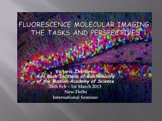
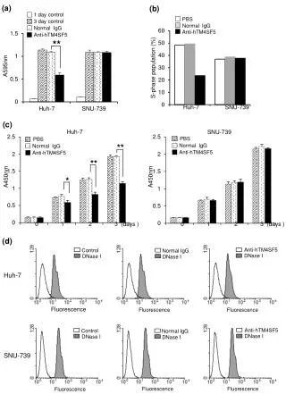
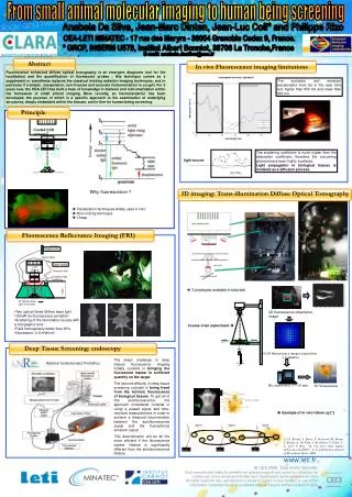
Cooled CCD camera. IAB. IR filter. Halogen lamp. Edges detection. 2D fluorescence reflectance image. Nodules in the lungs. Strong water attenuation. Strong heamoglobin attenuation. lens. Optical fibers. Laser source. liver. CCD Camera. Emission filter. Excitation light scattering.

E N D
Cooled CCD camera IAB IR filter Halogen lamp Edges detection 2D fluorescence reflectance image Nodules in the lungs Strong water attenuation Strong heamoglobin attenuation lens Optical fibers Laser source liver CCD Camera Emission filter Excitation light scattering day 10 day 12 day 14 ~20cm Why fluorescence ? IR filtered visible light illumination Visualization techniques widely used in vitro Non ionizing technique Cheap Reconstruction -> 5-10 min 3D Visualisation Abstract Principle In vivo Fluorescence imaging limitations The excitation and emission wavelengths must be in the near infra red: higher than 650 nm and lower than 900 nm. The scattering coefficient is much higher than the absorption coefficient, therefore the outcoming photons have been highly scattered. Light propagation in biological tissues is modeled as a diffusion process. light source 3D imaging: Trans-illumination Diffuse Optical Tomography Fluorescence Reflectance Imaging (FRI) µs>>µa • Two optical fibred 690nm laser light 100mW for fluorescence excitation • Scattering of the illumination source with a holographic lens • Field homogeneity better than 30% • Illumination: 2,6 mW/cm² IAB Deep Tissue Screening: endoscopy 10x10 fluorescence images acquisition -> 5 minutes IAB From small animal molecular imaging to human being screening Anabela Da Silva, Jean-Marc Dinten, Jean-Luc Coll* and Philippe Rizo CEA-LETI MINATEC - 17 rue des Maryrs - 38054 Grenoble Cedex 9, France. * GRCP, INSERM U578, Institut Albert Bonniot, 38706 La Tronche,France E-mail: anabela.dasilva@cea.fr Fluorescence enhanced diffuse optical tomography is an emergent diagnosis tool for the localization and the quantification of fluorescent probes ; this technique comes as a supplement or sometimes replaces the classical ionizing radiation imaging techniques, and in particular if a simple , inexpensive, non invasive and accurate instrumentation is sought. For 5 years now, the CEA-LETI has built a base of knowledge in markers and instrumentation within the framework of small animal imaging. More recently, an instrumentation has been developed, the purpose of which is a specific approach to the examination of underlying structures, deeply embedded within the tissues, and in fine for human being screening. Course of an experiment The major challenge in deep tissues fluorescence imaging initially consists in bringing the fluorescent marker in sufficient quantity on the target. The second difficulty in deep tissue screening consists in being freed from the intrinsic fluorescence of biological tissues. To get rid of this autofluorescence, the approach considered consists in using a pulsed signal and time-resolved measurements in order to achieve a temporal discrimination between the autofluorescence signal and the fluorophores emission signal. This discrimination will be all the more efficient if the fluorescence marker lifetime is significantly different from the autofluorescence lifetime. National funded project Prostafluo Exemple of in vivo follow up [*] [*] A. Koenig, L. Hervé, V. Josserand, M. Berger, J. Boutet, A. Da Silva, J.-M. Dinten, P. Peltié, J.-L. Coll, P. Rizo, “In vivo mice lungs tumors follow-up with fDOT”, to be published in Journal of Biomedical Optics 2008