Download

1 / 38
520 likes | 1.88k Vues
TNM staging and prognosis. Alexandru Eniu, MD, PhD Medical Oncologist Department of Breast Tumors Cancer Institute Ion Chiricu ţă Cluj-Napoca, Romania. The Basics of TNM Staging. Premises:

E N D
TNM staging and prognosis Alexandru Eniu, MD, PhD Medical Oncologist Department of Breast Tumors Cancer Institute Ion Chiricuţă Cluj-Napoca, Romania
The Basics of TNM Staging • Premises: • Cancers of the same anatomic site and histology share similar patterns of growth and similar outcomes. • As the size of the primary tumor (T) increases, regional lymph node involvement (N) and/or distant metastases (M) become more likely.
Diagnosis • THE ONLY CERTITUDE = PATHOLOGY • Always necessary • Insufficient for planning treatment • We need • prognostic factors • predictive factors • targeted diagnosis
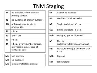
The Basics of TNM Staging • TNM records the 3 significant events in the life history of a cancer: • Local Tumor Growth (T) • TX, Tis, T0, T1, T2, T3, T4 • Spread to Regional Lymph Nodes (N) • NX, N0, N1, N2, N3 • Distant Metastasis (M) • MX, M0, M1
The Basics of TNM Staging • Stage Grouping • After assignment of TNM categories • Stage 0, I, II, III or IV • Multiple Simultaneous Tumors • The tumor with the highest T category is the one selected for classification and staging • Simultaneous bilateral cancers in paired organs are staged separately • Staging of primary unknown tumors can be based on clinical suspicion of the primary origin
Why Use TNM? • Allows the health professional to determine appropriate treatment ( primary, adjuvant) • Allows assessment of prognosis and outcomes • Enables the reliable evaluation of treatment results • Results in quality cancer care • Enables comparison of results
Primary Tumor (T) • Same definitions for clinical and pathologic T • If the measurement is made by physical examination, the examiner will use the major headings (T1, T2, or T3). • If mammographic or pathologic measurements are used, the subsets of T1 can be used. Tumors should be measured to the nearest 0.1 cm increment. • TX Primary tumor cannot be assessed • T0 No evidence of primary tumor • Tis Carcinoma in situ (DCIS, LCIS, Paget’s) • Note:Paget’s disease associated with a tumor is classified according to the size of the tumor.
Primary Tumor (T)Subdivisions of T1 • T1 Tumor 2 cm or less in greatest dim. • T1mic Microinvasion 0.1 cm or less in greatest dimension • T1a Tumor more than 0.1 cm but not more than 0.5 cm in greatest dimension • T1b Tumor more than 0.5 cm but not more than 1 cm in greatest dimension • T1c Tumor more than 1 cm but not more than 2 cm in greatest dimension
T2 Tumor more than 2 cm but not more than 5 cm in greatest dimension
T4Tumor of any size with direct extension to (a) chest wall or (b) skinT4a Extension to chest wall, not including pectoralis muscleT4bEdema (including peaud’orange) or ulceration of the skin of the breast, or satellite skin nodules confined to the same breastT4c Both T4a and T4bT4dInflammatory carcinoma
Inflammatory carcinoma vsneglected T4b T4d T4d T4b
Regional Lymph Nodes (N)Clinical • NX Regional lymph nodes cannot be assessed (e.g., previously removed) • N0Noregional lymph node metastasis • N1 Metastasis to movable ipsilateralaxillary lymph node(s) • N2 Metastases in ipsilateralaxillary lymph nodes fixed or matted, or in clinically apparent* ipsilateral internal mammary nodes in the absence of clinically evident axillary lymph node metastasis • N2a Metastasis in ipsilateralaxillary lymph nodes fixed to one another (matted) or to other structures • N2b Metastasis only in clinically apparent* ipsilateral internal mammary nodes and in the absence of clinically evident axillary lymph node metastasis Clinically apparent isdefined as detected by imaging studies (excluding lymphoscintigraphy) or by clinical examination or grossly visible pathologically.
Regional Lymph Nodes (N)Clinical • N3 Metastasis in ipsilateral infraclavicular lymph node(s) with or without axillary lymph node involvement, or in clinically apparent* ipsilateral internal mammary lymph node(s) and in the presence of clinically evident axillary lymph node metastasis; or metastasis in ipsilateral supraclavicular lymph node(s) with or without axillary or internal mammary lymph node involvement • N3a Metastasis in ipsilateral infraclavicular lymph node(s) • N3b Metastasis in ipsilateral internal mammary lymphnode(s) and axillary lymph node(s) • N3c Metastasis in ipsilateral supraclavicular lymph node(s)*Clinically apparent isdefined as detected by imaging studies (excluding lymphoscintigraphy) or by clinical examination or grossly visible pathologically.
Regional Lymph Nodes (N)Pathologic (pN) • pNX Regional lymph nodes cannot be assessed (e.g., previously removed, or not removed for pathologic study) • pN0No regional lymph node metastasis histologically, no additional examination for isolated tumor cells (ITC) • pN1 Metastasis in 1 to 3 axillary lymph nodes, and/or in internal mammary nodes with microscopic disease etected by sentinel lymph node dissection but not clinically apparent** • pN2 Metastasis in 4 to 9 axillary lymph nodes, or in clinically apparent* internal mammary lymph nodes in the absence of axillary lymph node metastasis • pN3 Metastasis in 10 or more axillary lymph nodes, or in infraclavicular lymph nodes, or in clinically apparent* ipsilateral internal mammary lymph nodes; or in ipsilateral supraclavicular lymph nodes
Distant Metastasis (M) • MX Distant metastasis cannot be assessed • M0 No distant metastasis • M1 Distant metastasis
Breast Cancer StagingStage I Stage 1 N0 T1
Breast Cancer StagingStage IIA Stage IIA N1 N0 N1 T2 T1 Stage IIa may also describe cancer in the axillary lymph nodes with no evidence of a tumor in the breast
Breast Cancer StagingStage IIB Stage IIB N1 N0 N1 T3 T2
Breast Cancer StagingStage IIIA Stage IIIA N2 N1 N1 T3 T1-3 N2 T1-3
N2 N2 T4 Breast Cancer StagingStage IIIB, IIIC Stage IIIB N0 N1 N1 T4 T4 Stage IIIC N3
Stage IV Breast Cancer • Stage IV breast cancer can be any size and has spread to distant sites in the body, usually the bones, lungs or liver, or chest wall
Breast Cancer Survival Rates Stage 2yr 5yr% 10yr% %BC I 100 90 70 60 II 90 70 55 30 III 70 40 30 IV (MBC) 25 2-5 <1 10 The overall median survival for MBC is <2ys. 50% of women with MBC stage IV will live <2ys.
How to Implement AJCC TNM Staging • Development of policy and procedure • Staging form part of the medical record • Development of a process by which the staging form is placed in the medical record • Size of facility and number of analytic cases • Pathology, medical records, cancer registry… • Development of Quality Control Methods to assure compliance
The TNM is imperfect! • Prognostic factors • Lymph Node Involvement • Tumor Size • Tumor Grade • Lymphatic/Vascular/Perineural Invasion • Age of the patient • Tumor biology Profile * ER, PR *Her2neu expression *Ki 67/ proliferation fraction
Future of Oncology • Diagnostic: Organ Molecular Etiology • Classification: Histology Molecular Function • Focus: Therapy Prevention • Therapy: Toxic, Complex Non-Toxic, Targeted • Outcome prediction: Suboptimal Precise • Patients follow-up : Anatomic Systemic
Regional Lymph Nodes (N) Pathologic (pN) a • pN0(i–) No regional lymph node metastasis histologically, negative IHC • pN0(i+) No regional lymph node metastasis histologically, positive IHC, no IHC cluster greater than 0.2 mm • pN0(mol–) No regional lymph node metastasis histologically, negative molecular findings (RTPCR)b • pN0(mol+) No regional lymph node metastasis histologically, positive molecular findings (RTPCR)b
Regional Lymph Nodes (N) • a Classification is based on axillary lymph node dissection with or without sentinel lymph node dissection. Classification based solely on sentinel lymph node dissection without subsequent axillary lymph node dissection is designated (sn) for “sentinel node,” e.g., pN0(i+) (sn). • b RT-PCR: reverse transcriptase/polymerase chain reaction.
Regional Lymph Nodes (N) • pN1 Metastasis in 1 to 3 axillary lymph nodes, and/or in internal mammary nodes with microscopic disease detected by sentinel lymph node dissection but not clinically apparent** • pN1mi Micrometastasis (greater than 0.2 mm, none greater than 2.0 mm) • pN1a Metastasis in 1 to 3 axillary lymph nodes • pN1b Metastasis in internal mammary nodes with microscopic disease detected by sentinel lymph node dissection but not clinically apparent** • pN1c Metastasis in 1 to 3 axillary lymph nodes and in internal mammary lymph nodes with microscopic disease detected by sentinel lymph node dissection but not clinically apparent.** (If associated with greater than 3 positive axillary lymph nodes, the internal mammary nodes are classified as pN3b to reflect increased tumor burden)
Regional Lymph Nodes (N) Pathologic (pN) a • pN2 Metastasis in 4 to 9 axillary lymph nodes, or in clinically apparent* internal mammary lymph nodes in the absence of axillary lymph node metastasis • pN2a Metastasis in 4 to 9 axillary lymph nodes (at least one tumor deposit greater than 2.0 mm) • pN2b Metastasis in clinically apparent* internal mammary lymph nodes in the absence of axillary lymph node metastasis • pN3 Metastasis in 10 or more axillary lymph nodes, or in infraclavicular lymph nodes, or in clinically apparent* ipsilateral internal mammary lymph nodes in the presence of 1 or more positive axillary lymph nodes; or in more than 3 axillary lymph nodes with clinically negative microscopic metastasis in internal mammary lymph nodes; or in ipsilateral supraclavicular lymph nodes
Regional Lymph Nodes (N) Pathologic (pN) a • pN3a Metastasis in 10 or more axillary lymph nodes (at least one tumor deposit greater than 2.0 mm), or metastasis to the infraclavicular lymph nodes • pN3b Metastasis in clinically apparent* ipsilateral internal mammary lymph nodes in the presence of 1 or more positive axillary lymph nodes; or in more than 3 axillary lymph nodes and in internal mammary lymph nodes with microscopic disease detected by sentinel lymph node dissection but not clinically apparent** • pN3c Metastasis in ipsilateral supraclavicular lymph nodes • *Clinically apparent is defined as detected by imaging studies (excluding lymphoscintigraphy) or by clinical examination. • **Not clinically apparent is defined as not detected by imaging studies (excluding lymphoscintigraphy) or by clinical examination.
Stage Grouping • Stage 0 Tis N0 M0 • Stage I T1* N0 M0 • Stage IIA T0 N1 M0 • T1* N1 M0 • T2 N0 M0 • Stage IIB T2 N1 M0 • T3 N0 M0 • Stage IIIA T0 N2 M0 • T1* N2 M0 • T2 N2 M0 • T3 N1 M0 • T3 N2 M0 • Stage IIIB T4 N0 M0 • T4 N1 M0 • T4 N2 M0 • Stage IIIC Any T N3 M0 • Stage IV Any T Any N M1 • *T1 includes T1mic • Note:Stage designation may be changed if post-surgical imaging studies reveal the presence of distant metastases, provided that the studies are carried out within 4 months of diagnosis in the absence of disease progression and provided that the patient has not received neoadjuvant therapy.
Schematic Diagram of Breast and Regional Lymph Nodes AJCC Cancer Staging Atlas