Download
1 / 1
10 likes | 145 Vues
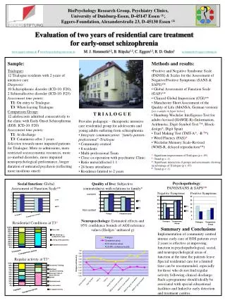
In vivo cardiac imaging system of zebrafish using a fluorescent dye for the assessment of anticancer drug-induced cardiotoxicity . . Short axis.

E N D
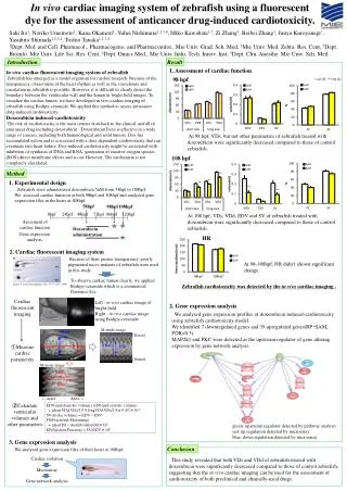
In vivo cardiac imaging system of zebrafish using a fluorescent dye for the assessment of anticancer drug-induced cardiotoxicity. Short axis Saki Ito1, Noriko Umemoto1, Kana Okamori1, YuheiNishimura1,2,3,4, MikoKawabata1,5, Zi Zhang1, BeibeiZhang1, JunyaKuroyanagi1, YasuhitoShimada1,2,3,4, Toshio Tanaka1,2,3,4 1Dept. Mol. and Cell. Pharmacol., Pharmacogeno. and Pharmacoinfor., Mie Univ. Grad. Sch. Med, 2Mie Univ. Med. Zebra. Res. Cent, 3Dept. Bioinfo. Mie Univ. Life Sci. Res. Cent, 4Dept. Omics Med., Mie Univ. Indu. Tech. Innov. Inst, 5Dept. Clin. Anesthe. Mie Univ. Sch. Med VDd VDs long axis VDd Introduction Result In vivo cardiac fluorescent imaging system of zebrafish Zebrafishhas emerged as a model organism for cardiac research. Because of the transparency, observation of the heart rhythm as well as the vasculature and circulation in zebrafish is possible. However, it is difficult to clearly detect the boundary between the ventricular wall and the lumen in bright field images. To visualize the cardiac lumen, we have developed in vivo cardiac imaging of zebrafish using Bodipy-ceramide. We applied this method to assess anticancer drug-induced cardiotoxicity. Doxorubicin induced-cardiotoxicity The risk of cardiotoxicity is the most serious drawback to the clinical usefull of anticancer drug including doxorubicin. Doxorubicin(Dox) is effective in a wide range of cancers, including both hematological and solid tumors. Dox has accumulated action and is associated with a dose dependent-cardiotoxicity that can eventuate into heart failure. Dox-induced cardiotoxicitymight be associated with inhibition of synthesis of DNA and RNA, generation of reactive oxygen species (ROS),direct membrane effects and so on. However, The mechanism is not completely elucidated. VDs 1. Assessment ofcardiac function 98 hpf *<0.05, **<0.01 At 98 hpf, VDs, but not other parameters of zebrafish treated with doxorubicin were significantly decreased compared to those of control zebrafish. 108 hpf Method 1. Experimental design Zebrafish were administered doxorubicin 5uM from 58hpf to 108hpf. We assessed cardiac function at both 98hpf and 108hpf and analyzed gene expression files in the heart at 108hpf. 58hpf 98hpf 108hpf At 108 hpf, VDs, VDd, EDV and SV of zebrafish treated with doxorubicin were significantly decreased compared to those of control zebrafish. Asessment of cardiac function Doxorubicin administration 0hpf 24hpf 48hpf 72hpf 96hpf 120hpf Gene expression analysis HR 2. Cardiac fluorescent imaging system Because of their greater transparency, poorly pigmented nacre mutants of zebrafish were used in this study. At98-108hpf, HR didn’t shown significant change. To observe cardiac lumen clearly, we applied Bodipy-ceramide which is a commercial fluorence dye. (Lister J, et al. Development 126: 3757-3767, 1999) Zebrafishcardiotoxicity wasdetected by the in vivo cardiac imaging . Cardiac fluorescent imaging Left : in vivo cardiac image of bright-field Right : in vivo cardiac image using Bodipy-ceramide 2. Gene expression analysis We analysed gene expression profiles of doxorubicin induced-cardiotoxicity using zebrafishcardiotoxicity model. We identified 7 downregulated genes and 39 upregulated genes(RP・SAM, FDR<0.3). MAP2k1and PKC were detected as the upstream regulator of gene altering expression by gene network analysis. M-mode image ↑Dorsal ①Measure cardiac parameters ↓Ventral M-mode image ←Aorta Apex→ ②Calculate ventricular volumes and other parameters EDV(end-diastolicvolume), ESV(end-systolic volume) = (shortVDd,VDs/2)2×longVDd,VDs/2×π×4/3×10-6 SV(stroke volume) = EDV-ESV FS(Fractional Shortening) = (shortDd-shortDs)/shortDd×102 EF(Ejection Fraction) = SV/EDV×102 green: upatream regulator detected by pathway analysis red: up regulation detected by microarray blue: down regulation detected by microarray 3. Gene expression analysis Conclusion We analysed gene expression files of their heart at 108hpf. Cardiac isolation Microarray Gene network analysis This study revealed that both VDs and VDdof zebrafish treated with doxorubicin were significantly decreased compared to those of control zebrafish, suggesting that the in vivo cardiac imaging can be used for the assessment of cardiotoxicity of both preclinical and clinically-used drugs. cut