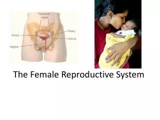
The Female Reproductive System
600 likes | 757 Vues
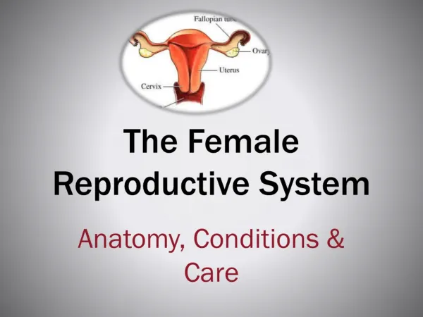
The Female Reproductive System. Blood Supply of Endometrium Arcuate arteries Encircle endometrium Radial arteries Supply straight arteries (to basilar zone) Supply spiral arteries (to functional zone). The Female Reproductive System. Cyclical Changes in Endometrium

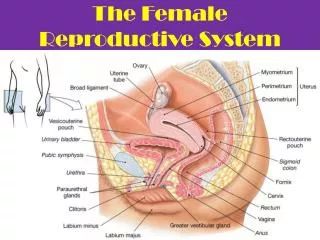
The Female Reproductive System
E N D
Presentation Transcript
The Female Reproductive System • Blood Supply of Endometrium • Arcuate arteries • Encircle endometrium • Radial arteries • Supply straight arteries (to basilar zone) • Supply spiral arteries (to functional zone)
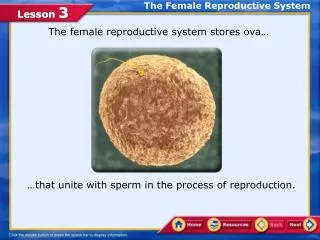
The Female Reproductive System • Cyclical Changes in Endometrium • Basilar zone remains relatively constant • Functional zone undergoes cyclical changes • In response to sex hormone levels • Produce characteristic features of uterine cycle
The Female Reproductive System • The Uterine Cycle (or menstrual cycle) • Is a repeating series of changes in endometrium • Lasts from 21 to 35 days • Average 28 days
The Female Reproductive System • Uterine Cycle • Responds to hormones of ovarian cycle • Menses and proliferative phase • Occur during ovarian follicular phase • Secretory phase • Occurs during ovarian luteal phase
The Female Reproductive System • Menses • Is the degeneration of functional zone • Occurs in patches • Is caused by constriction of spiral arteries • Reducing blood flow, oxygen, and nutrients • Weakened arterial walls rupture • Releasing blood into connective tissues of functional zone
The Female Reproductive System • Menses • Degenerating tissues break away, enter uterine lumen • Entire functional zone is lost • Through external os and vagina • Only functional zone is affected • Deeper, basilar zone is supplied by straight arteries
The Female Reproductive System Figure 26–20a The Appearance of the Endometrium during the Uterine Cycle.
The Female Reproductive System • Menstruation • Is the process of endometrial sloughing • Lasts 1–7 days • Sheds 35–50 mL (1.2-1.7 oz) blood
The Female Reproductive System • The Proliferative Phase • Epithelial cells of uterine glands • Multiply and spread across endometrial surface • Restore integrity of uterine epithelium • Further growth and vascularization • Completely restores functional zone • Occurs at same time as • Enlargement of primary and secondary follicles in ovary
The Female Reproductive System • The Proliferative Phase • Is stimulated and sustained by • Estrogens secreted by developing ovarian follicles • Entire functional zone is highly vascularized • Small arteries • Spiral toward inner surface • From larger arteries in myometrium
The Female Reproductive System Figure 26–20b The Appearance of the Endometrium during the Uterine Cycle.
The Female Reproductive System Figure 26–17 The Uterine Tubes.
The Female Reproductive System • Uterine Tube and Oocyte Transport • Involves ciliary movement and peristaltic contractions in walls of uterine tube • A few hours before ovulation, nerves from hypogastric plexus • “Turn on” beating pattern • Initiate peristalsis • From infundibulum to uterine cavity • Normally takes 3–4 days
The Female Reproductive System • Uterine Tube and Fertilization • For fertilization to occur • Secondary oocyte must meet spermatozoa during first 12–24 hours • Fertilization typically occurs • Near boundary between ampulla and isthmus
The Female Reproductive System • Uterine Tube and Nutrients • Uterine tube provides nutrient-rich environment • Containing lipids and glycogen • Nutrients supply spermatozoa and developing pre-embryo
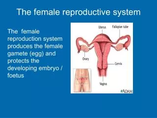
The Female Reproductive System • The Uterus • Provides for developing embryo (weeks 1–8) and fetus (week 9 through delivery): • Mechanical protection • Nutritional support • Waste removal
The Female Reproductive System • The Uterus • Is pear-shaped • 7.5 cm long, 5 cm diameter (3 in. x 2 in.) • Weighs 30–40 g (1-1.4 oz) • Normally bends anteriorly near base (anteflexion) • In retroflexion, uterus bends backward
The Female Reproductive System • Uterine Tube and Fertilization • For fertilization to occur • Secondary oocyte must meet spermatozoa during first 12–24 hours • Fertilization typically occurs • Near boundary between ampulla and isthmus
The Female Reproductive System • Uterine Tube and Nutrients • Uterine tube provides nutrient-rich environment • Containing lipids and glycogen • Nutrients supply spermatozoa and developing pre-embryo
The Female Reproductive System • The Uterus • Provides for developing embryo (weeks 1–8) and fetus (week 9 through delivery): • Mechanical protection • Nutritional support • Waste removal
The Female Reproductive System • The Uterus • Is pear-shaped • 7.5 cm long, 5 cm diameter (3 in. x 2 in.) • Weighs 30–40 g (1-1.4 oz) • Normally bends anteriorly near base (anteflexion) • In retroflexion, uterus bends backward
The Female Reproductive System • Three Suspensory Ligaments of Uterus • Uterosacral ligaments • Prevent inferior–anterior movement • Round ligaments • Restrict posterior movement • Cardinal(lateral) ligaments • Prevent inferior movement
The Female Reproductive System • Uterine Body (or corpus) • Is largest portion of uterus • Ends at isthmus • Fundus • Is rounded portion of uterine body • Superior to attachment of uterine tubes
The Female Reproductive System • Cervix • Is inferior portion of uterus • Extends from isthmus to vagina • Distal end projects about 1.25 cm (0.5 in.) into vagina • External os • Also called external orifice of uterus • Is surrounded by distal end of cervix • Leads into cervical canal
The Female Reproductive System • Cervical Canal • Is a constricted passageway opening to uterine cavity of body • At internal os (internal orifice)
The Female Reproductive System • Blood Supply of the Uterus • Branches of uterine arteries • Arising from branches of internal iliac arteries • Ovarian arteries • Arising from abdominal aorta • Veins and lymphatic vessels
The Female Reproductive System • Nerves of the Uterus • Autonomic fibers from hypogastric plexus (sympathetic) • Sacral segments S3 and S4 (parasympathetic) • Segmental blocks • Anesthetic procedure used during labor • Target spinal nerves T10–L1
The Female Reproductive System Figure 26–18a The Uterus.
The Female Reproductive System Figure 26–18b The Uterus.
The Female Reproductive System • The Uterine Wall • Has a thick, outer, muscular myometrium • Has a thin, inner, glandular endometrium (mucosa)
The Female Reproductive System • The Perimetrium • Is an incomplete serous membrane • Continuous with peritoneal lining • Covers • Fundus • Posterior surface of uterine body and isthmus
The Female Reproductive System • The Endometrium • Contributes about 10% of uterine mass • Glandular and vascular tissues support physiological demands of growing fetus • Uterine glands • Open onto endometrial surface • Extend deep into lamina propria
The Female Reproductive System Figure 26–19 The Uterine Wall.
The Female Reproductive System Figure 26–19 The Uterine Wall.
The Female Reproductive System • Estrogen • Causes uterine glands, blood vessels, and epithelium to change with phases of monthly uterine cycle
The Female Reproductive System • The Myometrium • The thickest portion of the uterine wall • Constitutes almost 90% of the mass of the uterus • Arranged into longitudinal, circular, and oblique layers • Provides force to move fetus out of uterus into vagina
The Female Reproductive System • Two Divisions of Endometrium • Functional zone • Layer closest to uterine cavity • Basilar zone • Adjacent to myometrium
The Female Reproductive System • The Functional Zone • Contains most of the uterine glands • Contributes most of endometrial thickness • Undergoes dramatic changes in thickness and structure during menstrual cycle
The Female Reproductive System • The Basilar Zone • Attaches endometrium to myometrium • Contains terminal branches of tubular endometrial glands
The Female Reproductive System • Blood Supply of Endometrium • Arcuate arteries • Encircle endometrium • Radial arteries • Supply straight arteries (to basilar zone) • Supply spiral arteries (to functional zone)
The Female Reproductive System • Cyclical Changes in Endometrium • Basilar zone remains relatively constant • Functional zone undergoes cyclical changes • In response to sex hormone levels • Produce characteristic features of uterine cycle
The Female Reproductive System • The Uterine Cycle (or menstrual cycle) • Is a repeating series of changes in endometrium • Lasts from 21 to 35 days • Average 28 days
The Female Reproductive System • Uterine Cycle • Responds to hormones of ovarian cycle • Menses and proliferative phase • Occur during ovarian follicular phase • Secretory phase • Occurs during ovarian luteal phase
The Female Reproductive System • Menses • Is the degeneration of functional zone • Occurs in patches • Is caused by constriction of spiral arteries • Reducing blood flow, oxygen, and nutrients • Weakened arterial walls rupture • Releasing blood into connective tissues of functional zone
The Female Reproductive System • Menses • Degenerating tissues break away, enter uterine lumen • Entire functional zone is lost • Through external os and vagina • Only functional zone is affected • Deeper, basilar zone is supplied by straight arteries
The Female Reproductive System Figure 26–20a The Appearance of the Endometrium during the Uterine Cycle.
The Female Reproductive System Figure 26–20b The Appearance of the Endometrium during the Uterine Cycle.
The Female Reproductive System Figure 26–17 The Uterine Tubes.
The Female Reproductive System • Uterine Tube and Oocyte Transport • Involves ciliary movement and peristaltic contractions in walls of uterine tube • A few hours before ovulation, nerves from hypogastric plexus • “Turn on” beating pattern • Initiate peristalsis • From infundibulum to uterine cavity • Normally takes 3–4 days