The Female Reproductive System
170 likes | 377 Vues
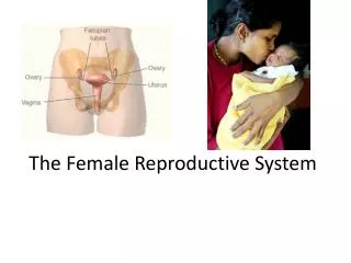
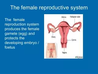
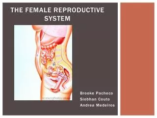
The Female Reproductive System. Brooke Pacheco Siobhan Couto Andrea Medeiros. Function and purpose. The female reproductive system produces sex cells and hormones. Protects and supports a developing embryo and provides the necessary nutrients to nourish an infant. Uterus.

The Female Reproductive System
E N D
Presentation Transcript
The Female Reproductive System Brooke Pacheco Siobhan Couto Andrea Medeiros
Function and purpose • The female reproductive system produces sex cells and hormones. • Protects and supports a developing embryo and provides the necessary nutrients to nourish an infant.
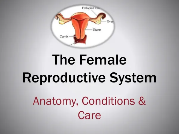
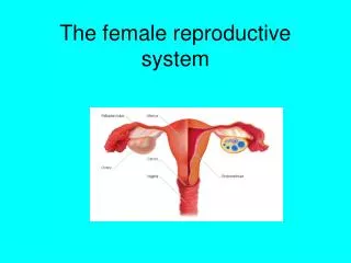
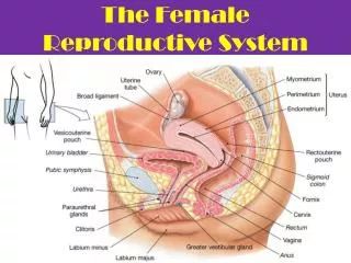
Uterus • A muscular chamber that provides mechanical protection and nutritional support for a developing embryo and fetus. • Small, pear-shaped organ • Uterus is split into the body and the cervix. The body ends in a constriction called the isthmus while the cervix is tubular and extends down to the vagina. • The uterine wall is made up of an inner endometrium which includes the epithelium lining the uterine cavity and the underlying connective tissues. • Muscular myometrium also makes up the lining and is found to be thickest in adult women who have not given birth
Vagina • An elastic, muscular tube extending between the uterus and the vestibule, a space bounded by the external genitalia. • The vagina lies parallel to the rectum and runs along the urethra. Three Major Functions; 1. Passageway for elimination of menstrual fluids 2.Receives the penis during sexual intercourse and holds spermatozoa prior to their passage into the uterus. 3.Forms lower portion of the birth canal which fetus passes during delivery. • Contains resident bacteria supported by nutrients in the cervical mucus. The metabolic activity of these bacteria create an acidic environment, restricting the growth of many pathogens
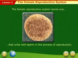
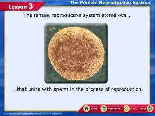
ovaries • Small, lumpy, oval shaped organs on either side of the pelvic cavity • Responsible for the production of female gametes, secretion of female sex hormones and the secretion of inhibin • 5cm x 2.5cm x 8mm • Pale white/yellowish coloring • Stabilized by the broad ligament and supporting ligaments
Ovarian Cycle • 28 day cycle that includes the follicular phase, ovulation and the luteal phase. • Follicular phase: begins on the first day of menstruation. Levels of estrogen and progesterone are at their lowest. Later in the follicular phase, proliferation (or thickening) of the uterine lining occurs. The follicular phase typically lasts about 14 days • Luteal Phase: Estrogen and progesterone increase, and work together to create changes in the lining of the uterus that prepare it to accept an embryo, should conception occur. When conception (or pregnancy) does not occur, estrogen and progesterone levels decline, and the lining of the uterus, or the endometrial lining, begins to shed, which leads to menstruation. • Ovulation: Ovulation is the release of a mature egg (ovum) from the ovarian follicle. Several ovarian follicles begin to mature and develop under the influence of pituitary hormones. Only one follicle develops fully. This dominant follicle produces an egg which will be released and can be fertilized. The follicle secretes increasing amounts of the hormone estrogen. Peak estrogen produces a surge of luteinizing hormone (LH).
oogenesis • Oogenesis is the ovum production – it begins before birth, accelerates during puberty and ends at menopause • Occurs every month during the ovarian cycle • Oogonia are the stem cells that complete their mitotic divisions in the ovaries before the female is even born. They make their way all the way to the prophase stage of meiosis I, but then stop. • They remain in that state until the female reaches puberty where a hormonal signal to complete meiosis is released. • 2 million oocytes are in the ovaries at birth, but only 400,000 remain at the start of puberty. • Ovarie follicles: specialized structures where oocyte growth and meiosis I of oogenesis occur.
Uterine tubes • Infundibulum: an expanded funnel at the end closest to the ovary with numerous fimbriae extend into the pelvic cavity. (both are covered with cilia) • The movement of that cilia, along with contractions of the smooth muscle of the wall transport the secondary oocyte. • It takes roughly 3-4 days for the oocyte to travel from the infundibulum to the uterine cavity. Unfertilized oocytes degenerate. • *If fertilization is to occur, the oocyte must encounter spermatozoa in the first 12-24 hours
Uterine cycle • A series of changes in the structure of the endometrium • Begins with the menarche, at age 11-12, and continues until menopause at age 45-55 • The time between the menarche and menopause – regular appearance of menstrual cycles. • Menstrual cycles are only interrupted by: illness, stress, starvation or pregnancy. • Occurs in three stages: menses, proliferative phase and secretory phase
menses • The degeneration of the superficial functional zone of the endometrium. It begins when there is a decrease in progesterone and estrogen levels. • The endometrial arteries constrict, reducing blood flow to the region. • Cells and tissues die of oxygen and nutrient deprivation. • Weakened arterial walls rupture and blood pours into the connective tissue of the functional zone. • When the blood enters the uterine cavity and is lost into the vagina, menstruation occurs. It generally takes 1-7 days and 35-50mL of blood is lost.
Proliferative phase • Begins within a few days of the end of menses. • The epithelial cells that survived multiply and spread across the surface of the endometrium. • This repair process occurs because of the increase in estrogen levels.
Secretory phase • The uterine glands enlarge, increasing rates of secretion as the endometrium prepares for the arrival of a developing embryo. • Stimulated by progestins and estrogens from the corpus luteum. • Begins at time of ovulation and lasts as long as corpus luteum is in tact.
External Genitalia • At the entrance of the vagina is the vestibule which includes all of the external genitalia. • Clitoris: a projection into the vestibule which engourges with blood during sexual arousal. Made up of the same embryonic structures as the male penis. • During arousal the lesser vestibular glands discharge secretions to keep the vestibule moist while the greater vestibular glands discharge secretions near the vaginal entrance.
Mammary Glands • In females, mammary glands are specialized organs organs of the integumentary system and controlled by hormones of he reproductive system. • The ducts of the underlying mammary gland open onto the body through the nipple. • Each gland is made up of a number of different lobes containing milk glands. • Mammary glands are necessary for the development of infants through lactation.
Endometriosis • Endometriosis is a female health disorder where the lining of the uteran walls grow in other areas of the body and shed. • Cause: Unknown • Symptoms: Painful periods, pain during intercourse, pain with bowel movements and lower back pain during menstrual cycle. • Treatment: Medications to control pain, hormone medications to stop the endometriosis from getting worse, or surgery to remove the areas of endometriosis or the entire uterus and ovaries • Complications: Can lead to problems when trying to get pregnant.
Works Cited • Board, A.D.A.M. Editorial. "Causes, Incidence, and Risk Factors." Endometriosis. U.S. National Library of Medicine, 18 Nov. 0000. Web. 29 Apr. 2012. <http://www.ncbi.nlm.nih.gov/pubmedhealth/PMH0001913/>. • Martini, Frederic, and Edwin F. Bartholomew. Essentials of Anatomy and Physiology. San Francisco: Benjamin Cummings, 2009. Print. • "The Leading Provider of Science and Specialist Images." Science Photo Library. Web. 29 Apr. 2012. <http://www.sciencephoto.com/>. • "Ovulation." Ovulation Calendar and Ovulation Chart. Web. 29 Apr. 2012. <http://www.fertilityfriend.com/Faqs/Ovulation.html%20>.