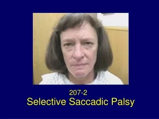
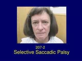
207-2
207-2. Selective Saccadic Palsy. Selective Saccadic Palsy after Cardiac Surgery. Selective loss of all forms of saccades (voluntary and reflexive quick phases of nystagmus) with sparing of other eye movements. Patterns of Saccadic Movements.

207-2
E N D
Presentation Transcript
207-2 Selective Saccadic Palsy
Selective Saccadic Palsy after Cardiac Surgery Selective loss of all forms of saccades (voluntary and reflexive quick phases of nystagmus) with sparing of other eye movements.
Patterns of Saccadic Movements Slow saccades that carry the eye almost to the target. A “staircase” of 10 or more small saccades, to acquire the target. *Seen clinically like a slow smooth movement Hypometric saccades combined with slowing. Loss of all ability to make saccades and reflexive quick phases.
Patterns of Saccadic Movements Slow horizontal and vertical saccades 9/10 Slow vertical saccades only 1/10 Slow horizontal saccades only – not present Solomon D et al., Ann Neurol 2007; 62: 1-11
Figure 1. Examples of horizontal saccadic abnormalities after cardiac surgery. (A) Accurate saccade made by a healthy subject. (B) Slow saccade made by Patient 9 (P9) that is slightly hypometric. (C) Slow saccade made by P1 that is hypometric; note that velocity dips and then increases (arrow), suggesting more than one pulse of innervation. (D) Pronounced hypometria of saccades made by P4, with a staircase of small movements that take the eye to its target. Positive values indicate rightward movements. Note that scales differ for each panel. Red lines designate eye position; dashed lines designate target position; blue lines designate eye velocity.
Figure 2. Representative examples of preservation of other types of eye movements in Patient 3 (P3), who had both horizontal and vertical saccadic palsy, and P10, who had complete saccadic palsy. (A) Example of tonic deviation of the eyes in the direction of upward optokinetic stimulus motion in P3; resetting quick phases are small. Red line indicates vertical gaze; blue line indicates horizontal gaze. (B) Convergence movement of about 15 degrees (positive value) in P3, made with a small upward saccade. Red line indicates vertical gaze; blue line indicates vergence. (C) Horizontal vestibuloocular reflex during passive yaw head rotation as P3 looked toward a flashing light in a dark room; gain is about 1.0, so that gaze (eye position in space) remains almost constant. Green line indicates head; blue line indicates gaze; red line indicates eye in head. (D) Onset and subsequent horizontal smooth pursuit in P10; note how he generates smooth movements with little phase shift compared with the target (dashed line), despite complete absence of corrective saccades. Red line indicates horizontal gaze.
Figure 3. Schematic of brainstem components of saccade-generating mechanism, with hypothetical sites at which slow or hypometric saccades might arise. Excitatory burst neurons (EBNs) receive a trigger signal from the superior colliculus (not shown), which is relayed by long-lead burst neurons (LLBNs) and uses glutamatergic mechanisms. The second major projection to burst neurons is from omnipause neurons (OPNs), which are tonically active but are inhibited by the superior colliculus when a saccade is to be generated. OPNs inhibit burst neurons via glycine. When a saccade is to be triggered, OPNs cease discharge, releasing burst neurons from inhibition. The trigger signal is amplified by glycine, which also acts as a neuromodulator at glutamatergic receptors. OPN neurons cease firing during the saccade but resume when a motor error signal falls to near zero, signaling that the saccade is complete. Slow saccades could be caused by (1) lesions affecting EBN, (2) lesions affecting OPNs, or (3) an abnormal trigger signal. Hypometric saccades might arise if the threshold at which the motor error signal allows OPNs to resume discharge is increased (4). MN = motoneuron.
References Hanson MR, Hamid MA, Tomsak RL, Chou SS, Leigh RJ. Selective saccadic palsy caused by pontine lesions: clinical, physiological, and pathological correlations. Ann Neurol 1986;20(2):209-217. Tomsak RL, Volpe BT, Stahl JS, Leigh RJ. Saccadic palsy after cardiac surgery: visual disability and rehabilitation. Ann N Y Acad Sci 2002;956:430-433.
Solomon, D, Ramat S, Tomsak RL, Reich SG, Shin RK. Zee DS, Leigh RJ. Saccadic Palsy following Cardiac Surgery: Characteristics and Pathogenesis. Ann of Neurol 2007;62:1-11. Mokri B, Ahlskog JE, Fulgham JR, Matsumoto JY. Syndrome resembling PSP after surgical repair of ascending aorta dissection or aneurysm. Neurology 2004;62(6):971-973.
Acknowledgment Eggers SD, Moster ML, Cranmer K. Selective saccadic palsy following cardiac surgery. Neurology 2008;70:318-320.