Download
1 / 48
480 likes | 504 Vues
Dive into the intricate world of the nervous system, from the central to peripheral divisions, brain development, and the relationship between brain size and intelligence. Discover the complexities of the cortex, lobes, and functional areas, including the frontal lobes' role in movements and the prefrontal cortex's impact on behavior. Explore lobotomies, parietal lobe functions, and the importance of the temporal lobes. Unravel the mysteries of brain structure and function in this comprehensive guide to the nervous system.

E N D
The Central nervous system The peripheral nervous system Development and change in the nervous system The Functions of the Nervous System Chapter 3
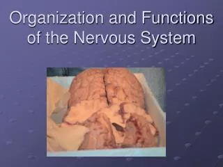
The Central Nervous System • The nervous system has two major divisions, the central nervous system (CNS) and the peripheral nervous system (PNS) • In the CNS (brain and spinal cord): • A bundle of axons traveling together is called atract. • A group of cell bodies is called anucleus. • In the PNS: • A bundle of axons travelling together is called anerve. (A neuron is not a nerve.) • A group of cell bodies is called aganglion. ◊
The Central Nervous System • During development, three major parts of the brain are formed: the forebrain, midbrain, and hindbrain. • The forebrain is the largest part of the brain. • It is made up of two cerebral hemispheres separated by the longitudinal fissure. • The cortex covers the cerebral hemispheres and is wrinkled or convoluted, increasing the amount of cortex. • A ridge is called a gyrus. • A groove is called a sulcus or, if large, a fissure. • The cortex is composed of layers and columns. • The forebrain also includes the thalamus and hypothalamus.
CNS Development Figure 3.3
Layers and Columns of the CortexFigure 3.6 The layered structure of the cortex. Columnar arrangement.
The Central Nervous System • Do intelligent people have bigger brains? • Brain size is mostly related to body size, because larger bodies require larger brains. • Examples: Elephants and sperm whales have brains that are 5-6 times larger than humans. • Among humans, there is a correlation between brain size and intelligence. • However, the relationship is small and highly variable. ◊
The Central Nervous System • Two key features characterize brains of more “intelligent” species. • The cortex has more convolutions. • The cerebral hemispheres are larger in proportion to the lower areas of the brain. • This illustrates ahierarchyof increasing complexity from spinal cord to hindbrain to midbrain and finally to the forebrain. ◊
The Central Nervous System • The four lobes of the brain: • Frontal lobe • In front of (anterior to) the central sulcus • Above (superior to) the lateral fissure • Parietal lobe • Behind (posterior to) the central sulcus • Occipital lobe • At the back (posterior) of the brain • Temporal lobe • Located on the sides (laterally) of the brain • These are somewhat arbitrary divisions but useful for locating structures and functions.
The Central Nervous System • The frontal lobes are important for movement and complex human capabilities. • The primary motor cortex • is found on theprecentral gyrus; • is a “map” of the human body, or homunculus, with larger areas devoted to parts of the body that make precise movements; • works with secondary motor cortex and with subcortical structures, for example, the basal ganglia. • Broca’s area is important for speech production. ◊
The Central Nervous System • Prefrontal cortex: • is the largest part of the human brain; • plays a role in organizing and planning; • is involved in some types of decision making; • is important for impulse control; • adjusts behavior in response to rewards and punishments. • Prefrontal lobe dysfunction: • impairs the ability to learn from consequences; • decreases the ability to control impulses; • is often found in depression and schizophrenia.
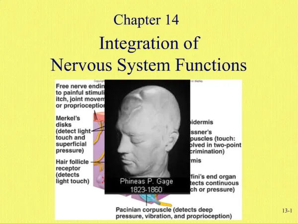
The Central Nervous System • Frontal lobotomy • is a surgical procedure that disconnects the prefrontal area from the rest of the brain; • was performed on 40,000 patients in the U.S. during the 1940s and 1950s, mostly to calm agitated patients; • provided little benefit at high cost to the patient; • has largely been replaced by drug treatment, along with other forms of psychosurgery; • occurred as the result of an accident in the famous case of Phineas Gage. ◊
The Central Nervous System • The parietal lobes are important for body sensations and spatial localization. • The primary somatosensory cortex • is located on the postcentral gyrus; • receives information about the skin senses, body position, and movement; • maps these functions as a sensory homunculus (with size corresponding to sensitivity in that part of the body). ◊
The Central Nervous System • Parietal association areas • combine information from body senses and vision; • identify objects by touch, determine the location of the limbs, and locate objects in space. • Damage to the posterior parietal cortex causes neglect of objects, people, and activity on the opposite side. • Usually this is caused by damage on the right side. • The patient may deny there is anything wrong, even when a limb is paralyzed. ◊
The Central Nervous System • The temporal lobes are separated from the frontal and parietal lobes by the lateral fissure; they: • contain the auditory cortex, which receives information from the ears; • include language and auditory and visual association areas. • Wernicke’s area is involved in language comprehension and production. • Damage results in meaningless speech and poor comprehension of written and spoken communication. • The inferotemporal cortex is concerned with visual identification. Damage causes difficulty in recognizing objects and familiar faces.
The Central Nervous System • The occipital lobes • are located in the posterior part of the brain; • are the location of the primary visual cortex; • contain a map of visual space because adjacent receptors in the eye send information to adjacent points in the visual cortex; • have secondary visual areas that process individual components of a scene, including • color, • movement, • and form.
The Central Nervous System • Another structure in the forebrain is the thalamus, located below the lateral ventricles. • It receives information from all the senses except olfaction. • It relays this sensory information to the cortex. • Some portions of the thalamus project more diffusely and play a role in arousal. • The hypothalamus is located beneath the thalamus. • It controls emotions and motivated behaviors such as eating, drinking, and sexual activity. • It exerts major control over the autonomic nervous system and the endocrine system (by way of the pituitary).
The Central Nervous System • Just posterior to the thalamus is the pineal gland. • The pineal gland secretes melatonin, a hormone that induces sleep. • It controls seasonal cycles in nonhuman animals . • It participates with other structures in controlling daily rhythms in humans. • The corpus callosum, a dense band of fibers at the bottom of the longitudinal fissure, shares information between the hemispheres. • Epilepsy patients whose corpus callosum has been severed have been helpful in studying the specializations of the two hemispheres.
The Central Nervous System • During development, the hollow interior of the nervous system becomes the ventricles of the brain and the central canal in the spinal cord. • The ventricles are filled with cerebrospinal fluid that • carries material from the blood vessels to the central nervous system • transports waste materials in the other direction. • The two lateral ventricles and the third ventricle are found in the forebrain. ◊
The Central Nervous System • The midbrain contains structures that have secondary roles in vision, audition and movement. • The superior colliculi help guide eye movements and fixation of gaze. • Theinferior colliculi help locate the direction of sounds. • Thesubstantia nigra projects to the basal ganglia to integrate movements. • Theventral tegmental areaplays a role in the rewarding effects of food, sex, drugs and so on. • The midbrain is located at the top of thebrain stem. ◊
The Central Nervous System • The hindbrain is composed of the medulla, the pons, and the cerebellum. • The medulla is involved in the control of essential life processes such as cardiovascular activity and respiration (breathing). • Theponscontains centers related to sleep and arousal, which are part of the reticular formation. • Thereticular formation contributes to attention, reflexes, and muscle tone. ◊
The Central Nervous System • The cerebellum, is the most distinctive structure in the brain stem. • It is located on the back of the brain stem. • It is wrinkled and divided down the middle like the cerebral hemispheres, and is often referred to as the “little brain.” • The cerebellum refines movements initiated by the motor cortex by controlling their speed, intensity and direction. • It also plays a role in motor learning, as well as in other cognitive processes and in emotion. ◊
The Central Nervous System • The spinal cord is a finger-sized cable of neurons that carries commands from the brain to the muscles and organs, and sensory information into the brain. • Sensory neurons enter the spinal cord through the dorsal root of each spinal nerve. • The axons of the motor neurons pass out through the ventral root. • In some cases sensory neurons connect, directly or through an interneuron, with motor neurons; this pathway produces a simple, automatic movement in response to a sensory stimulus, called a reflex. ◊
The Central Nervous System • Protecting the Central Nervous System • The space between the meningesand the CNS is filled with cerebrospinal fluid, which cushions the neural tissue from blows and sudden movement. • The blood-brain barrier limits passage into the brain of toxic substances and neurotransmitters circulating in the blood. • Some places are not protected by this barrier, such as the area postrema, which produces vomiting if toxins are ingested. ◊
The Peripheral Nervous System • The peripheral nervous system (PNS) • is made up of: • the cranial nerves that enter and leave the underside of the brain: • and the spinal nerves that connect to the sides of the spinal cord at each vertebra; • and can be divided into : • the somatic nervous system, composed of the motor and sensory neurons that allow us to sense and react to the environment; • the autonomic nervous system (ANS), which regulates general activity levels in the body and controls smooth muscle, the glands, the heart, and other organs.
Divisions of the Nervous System Figure 3.19
The Peripheral Nervous System • The autonomic nervous system is composed of two branches. • The sympathetic nervous system • activates the body in ways that help it cope with demands, such as emotional stress and physical emergencies; • has most of its ganglia in thesympathetic ganglion chain. • The parasympathetic nervous system • slows the activity of most organs to conserve energy; • activates digestion to renew energy; • has its ganglia near the muscles and glands they control. ◊
Development and Change in the Nervous System • The nervous system begins as a hollow tube that later becomes the brain and spinal cord. • The nervous system begins development when the surface of the embryo forms a groove. • The edges of this groove curl upward until they meet, turning the groove into a tube. • Further development occurs in four stages: • cell proliferation • migration • circuit formation • circuit pruning
Development and Change in the Nervous System • During proliferation the cells that will become neurons divide and multiply. • Occurs in the ventricular zone, the area surrounding the hollow tube. • 250,000 new cells are “born” every minute. • These newly formed neurons then migrate, moving from the ventricular zone outward to their final location. • They do so with the aid of specialized radial glial cells. • Each neuron’s function is determined by the time and location that it develops.
Development and Change in the Nervous System • During circuit formation, the axons of developing neurons grow toward their target cells and form functional connections. • Growth cones at the axon’s tip guide the migrating axons. • They detect chemical and molecular signposts that attract or repel the advancing axon. • Using these signposts, the axon is able to navigate to intermediate stations and past inappropriate targets until they reach their final destinations. ◊
Development and Change in the Nervous System • The route to the destination is not always direct. • Changing genetic controls allow the axon to make course changes along the way; for example: • While the gene Robo1 is active, a neuron is repelled from the brain’s midline by a chemical located there. • If the axon is to cross the midline, Robo3 becomes active at the appropriate place, and the axon is attracted to the midline. • After crossing, control reverts to Robo1 and the midline is avoided. ◊
Development and Change in the Nervous System • During circuit pruning excess neurons and synapses are eliminated. • Neurons that are unsuccessful in finding a place on a target cell or that arrive late die. • Next, the nervous system eliminates excessive synapses. • Synapses are strengthened or weakened depending on whether the presynaptic neuron and the postsynaptic neuron fire together. • Postsynaptic neurons apparently release neurotrophins that enhance development in the presynaptic neuron. • Later the plasticity (ability to be modified) of these synapses decreases.
Development and Change in the Nervous System • Fetal alcohol syndrome (FAS), which often produces mental retardation, is caused by the mother’s use of alcohol during a critical period of brain development. • FAS brains are often small and malformed, and neurons are dislocated. • During migration many cortical neurons fail to line up in columns as they normally would, because the radial glial cells revert to their more typical glial form prematurely. • Other neurons continue migrating beyond the usual boundary of the cortex. • Exposure to ionizing radiation affects both proliferation and migration.
Development and Change in the Nervous System • Stimulation continues to shape synaptic construction and reconstruction throughout the individual’s life. • Much of the change resulting from experience in the mature brain involves reorganization. • Reorganization is a shift in connections that changes the function of an area of the brain. • Reorganization may provide compensation for losses, for example, in syndactyly and blindness. • However, reorganization is not always beneficial. • Example: kittens reared in abnormal visual environments • Example: phantom pain
Development and Change in the Nervous System • Two major sources of brain injury are: • Stroke • Stroke is caused by artery blockage (ischemic) or rupture (hemorrhagic). • Damage is due to oxygen and glucose deprivation, excitotosis, and edema (swelling). • Stroke is a leading cause of death and disability in the U.S. • Traumatic Brain Injury • Traumatic brain injury is caused by a blow to the head, penetration, or sudden acceleration or deceleration. • Even trauma that does not produce concussion can result in brain changes typically seen in Alzheimer’s patients.
Development and Change in the Nervous System • Limitations of Self Repair • Regeneration is the regrowth of severed axons. • Myelin provides a guide tube for the neuron to grow through, and the axon is guided to its destination much as in development. • Occurs in the amphibian brain and in the mammalian PNS. • In the mammalian CNS, glia produce scar tissue and growth inhibitors, and immune cells may also interfere. ◊
Development and Change in the Nervous System • Neurogenesisis the birth of new neurons. • It appears to support learning (in the hippocampus) and odor discrimination (in the olfactory bulbs). • There is no evidence neurogenesis contributes to self repair. • However neurogenesis does increase in damaged brains, and there is some hope this could be enhanced as a means of recovery. ◊
Development and Change in the Nervous System • Compensation • Presynaptic neurons sprout more terminals to form additional synapses. • Postsynaptic neurons add more receptors. • Silent side branches from adjacent neurons become active within minutes of injury. • Reorganization • Functions are taken over by other areas. • Typically, compensation is by an adjacent area, but it can involve the other hemisphere. • Reorganization is more likely if the damage occurs early in life.
Development and Change in the Nervous System • Possibilities for CNS Repair • Neuron growth enhancers • Growth enhancer in brain-damaged rats induced axons to grow from the undamaged side into the spinal cord pathway on the other side; the rats recovered most of their mobility. • Providing guide tubes or scaffolding • Rats regained use of their legs when glial cells were provided as scaffolding between the cut ends of the spinal cord. • Almost no improvement occurred in trials with humans. • Counteracting regrowth inhibitors • Monkeys recovered 80% of the use of their paralyzed hands after the effects of Nogo-A (which inhibits axon growth) were blocked.
Development and Change in the Nervous System • Stem cells seem an ideal means of neural repair. • Stem cells are undifferentiated cells that can develop into specialized cells such as neurons, muscle, or blood. • Embryonic stem cells placed in an adult nervous system differentiate into neurons appropriate to that area. • Later in life, stem cells lose most of their flexibility, but those found in the olfactory mucosa show promise for repairing neural damage. ◊
Development and Change in the Nervous System • Other efforts at CNS repair utilize computer chips. • One sends signals from the brain directly to a paralyzed limb, bypassing the damaged area in the spinal cord. • Monkeys with a blocked nerve learned to flex, extend, and rotate the paralyzed wrist. • Another device simulates a central pattern generator in the spinal cord. • With just 10 simulated neurons and 190 synapses, it allowed a cat with paralyzed hind legs to walk. • A chip implanted in a paraplegic’s motor cortex enabled him to operate a robotic arm and to access email on a computer.