RNA SPLICING AND PROCESSING CHAPTER 21 ( GENES X )
1.16k likes | 1.61k Vues
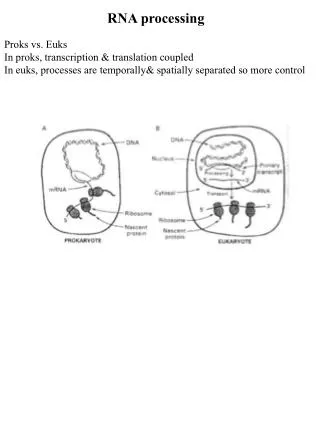
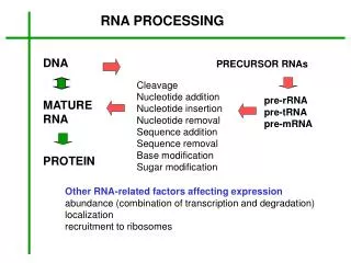
RNA SPLICING AND PROCESSING CHAPTER 21 ( GENES X ). Introduction to RNA processing. Transcription elongation is tightly coupled to RNA processing. Eukaryotic mRNAs are modified at their beginning, middle and end. Why modify mRNA? Assess if mRNA is intact

RNA SPLICING AND PROCESSING CHAPTER 21 ( GENES X )
E N D
Presentation Transcript
RNA SPLICING AND PROCESSING CHAPTER 21 (GENES X)
Introduction to RNA processing Transcription elongation is tightly coupled to RNA processing. Eukaryotic mRNAs are modified at their beginning, middle and end Why modify mRNA? Assess if mRNA is intact Provides a regulatory mechanism for the amount of protein produced by a gene Introns-different forms of a protein from the same gene
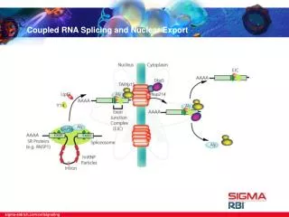
Introduction to RNA processing I. mRNA processing (eukaryotes) 1. 5´ capping 2. 3´ cleavage and polyadenylation 3. RNA splicing (nuclear pre-mRNA) II. Nuclear pore complex overview III. rRNA and tRNA processing overview
mRNA processing • Occurs in eukaryotes (but a few bacterial cases exist) • Primary RNA polymerase II transcripts (pre-mRNA) are usually not functional • RNA is processed following (or during) transcription
3 steps of mRNA processing occurs in the nucleus 5’ CAP Poly-A tail Splicing
CAP- THIS IS the 5’ end of an mRNA! Marks mRNA for export to cytoplasm for translation
The capping reaction is performed by three enzymes acting in succession: • A phosphatase: removes one phosphate from the 5’ end of the nacent RNA, • A guanyl transferase: adds GMP in a reverse linkage (5’ to 5’ instead of 5’ to 3’), • A methyl transferase: adds the methly group to the guanosine.
5´ capping • 5´ end of pre-mRNA is covalently modified • 7-methylguanosine is added • Linked 5´ to 5´ • Occurs shortly after initiation
Function of 5´ cap • Protection from degradation • Increased translational efficiency • Transport to cytoplasm • Splicing of first exon
Do all RNA polymerases CAP? Pol I and III do not have the CTD, and do not cap their RNAs The cap distinguishes mRNAs
Structure of two human genes showing the arrangement of exons and introns. (A) The relatively small b-globin gene, which encodes one of the subunits of the oxygen-carrying protein hemoglobin, contains 3 exons (see also Figure 4–7). (B) The much larger Factor VIII gene contains 26 exons; it codes for a protein (Factor VIII) that functions
Variation in intron and exon lengths in the human, worm, and fly genomes. Size distribution of exons. (B) Size distribution of introns. Note that exon length is much more uniform than intron length.
Introduction to RNA processing I. mRNA processing (eukaryotes) 3. RNA splicing (nuclear pre-mRNA) A. Introns, exons, and splicing B. Spliceosome and snRNAs C. Self-splicing D. Alternative splicing
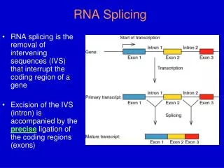
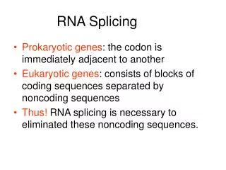
RNA Splicing • Primary transcripts (in eukaryotes) are sometimes “spliced” to remove non-coding regions “introns” from coding regions “exons” • The exon regions are spliced together to form the mature mRNA hnRNA addition of cap, polyA tail - Poly A Tail 5’ Cap- Splicing - Poly A Tail 5’ Cap- Mature mRNA
Types of mRNA Splicing • Types I & II: self-splicing of catalytic RNA sequences (Ribozymes) • - Two types reflect different ribozyme types • Type III: occurs in a protein - RNA complex - Responsible for nearly all splicing • - It has been speculated that the RNA component of this structure is the catalytic component which would also make it a ribozyme
Prevalence Of Splicing Type III: • - most mRNAs in vertebrates • - many mRNAs in invertebrates • - some mRNAs in unicellular • eukaryotes mRNA: Eukaryotes no type III mRNA splicing in Prokaryotes! Type I & II: Eukaryotes - rRNA in tetrahymena nuclei (Type I) - mRNA & rRNA in fungal mitochondria (I & II) - mRNA in some chloroplasts (II) Prokaryotes - mRNA in bacteriophage T4 (I)
Mechanism of pre-mRNA Splicing(Spliceosome Mediated) Type III • - Introns have conserved sequences at the splice junctions • - SnRNPs (Small nuclear ribonuclear proteins) binds critical sites on the pre-mRNA • - pronounced ‘SNURPs’ • - these are complexes containing both protein and small RNAs • the small RNAs are transcribed by RNA polymerase III • they then associate with accessory proteins • the complex then recognizes critical sites for splicing by base pairing
The consensus nucleotide sequences in an RNA molecule that signal the beginning and the end of most introns in humans. Only the three blocks of nucleotide sequences shown are required to remove an intron sequence; the rest of the intron can be occupied by any nucleotide. Here A, G, U, and C are the standard RNA nucleotides; R stands for either A or G; Y stands for either C or U. The A highlighted in red forms the branch point of the lariat produced by splicing. Only the GU at the start of the intron and the AG at its end are invariant nucleotides in the splicing consensus sequences. The remaining positions (even the branch point A) can be occupied by a variety of nucleotides, although the indicated nucleotides are preferred. The distances along the RNA between the three splicing consensus sequences are highly variable; however, the distance between the branch point and 3' splice junction is typically much shorter than that between the 5' splice junction and the branch point.
- consensus sequences are conserved throughout eukaryotes Conservation of sequence is expected with recognition of sequences being done by base pairing with snRNP’s RNA component
The RNA splicing reaction. (A) In the first step, a specific adenine nucleotide in the intron sequence (indicated in red) attacks the 5' splice site and cuts the sugar-phosphate backbone of the RNA at this point. The cut 5' end of the intron becomes covalently linked to the adenine nucleotide, as shown in detail in (B), thereby creating a loop in the RNA molecule. thereby creating a loop in the RNA molecule. The released free 3'-OH end of the exon sequence then reacts with the start of the next exon sequence, joining the two exons together and releasing the intron sequence in the shape of a lariat. The two exon sequences thereby become joined into a continuous coding sequence; the released intron sequence is degraded in due course.
Nuclear pre-mRNA splicing • Introns: non-coding sequences • Exons: coding sequences • RNA splicing: removal of introns and joining of exons • Splicing mechanism must be precise to maintain open reading frame • Catalyzed by spliceosome (RNA + protein)
Classes of intronic splicing: splicesome • Major pathway for splicing and is used for processing of all pre-mRNAs in eukaryotes • Splicesome is a complex of five small nuclear ribonucleoprotein particles (snRNPs). • Each snRNPs consists of a small nuclear RNAs (snRNA) associated specifically with proteins. • Large, ~60S (size of a ribosome), 5 snRNAs (U1, U2, U4, U5, U6) and roughly 50 proteins! • Splice at specific markers with the mRNA using the snRNA for recognition and catalysis!
Once all of the different snRNPs associate with their appropriate targets on the pre-mRNA the entire (very large) complex is called a: Spliceosome
Splicesomal mechanism • snRNA U1 has sequences that are complementary to 5’ splice site • U1 plus its protein component is the U1 snRNP • upon U1 snRNP binding, U2, U4, U5 and U6 snRNP assemble • ATP is required for assembly but not catalysis! (needed for RNA helicases that allow alternative pairing schemes) Proteins removed from this view for clarity!
Secondary structure model of human U1 snRNP. The region where it recognizes the pre-mRNA is also shown
The E complex contains: • U1 snRNP bound at the 5′ splice site • the protein U2AF bound to a pyrimidine tract between the branch site and the 3′ splice site • SR proteins connecting U1 snRNP to U2AF Important point is recognition and complementarity of 5’ splice site sequences with U1 RNA sequences. 35 Figure 26.10
Splicesomal mechanism ATP is required for assembly but not for catalysis
The RNA splicing mechanism. RNA splicing is catalyzed by an assembly of snRNPs (shown as colored circles) plus other proteins (most of which are not shown), which together constitute the spliceosome. The spliceosome recognizes the splicing signals on a pre-mRNA molecule, brings the two ends of the intron together, and provides the enzymatic activity for the two reaction steps (see Figure 6–26). The branch-point site is first recognized by the BBP (branch-point binding protein) and U2AF, a helper protein. In the next steps, the U2 snRNP displaces BBP and U2AF and forms base pairs with the branch- point site consensus sequence, and the U1 snRNP forms base-pairs with the 5' splice junction (see Figure 6–30). At this point, the U4/U6∑U5 “triple” snRNP enters the spliceosome.
In this triple snRNP, the U4 and U6 snRNAs are held firmly together by base-pair interactions and the U5 snRNP is more loosely associated. Several RNA–RNA rearrangements then occur that break apart the U4/U6 base pairs (as shown, the U4 snRNP is ejected from the splicesome before splicing is complete) and allow the U6 snRNP to displace U1 at the 5' splice junction (see Figure 6–30). Subsequent rearrangements create the active site of the spliceosome and position the appropriate portions of the pre-mRNA substrate for the splicing reaction to occur. Although not shown in the figure, each splicing event requires additional proteins, some of which hydrolyze ATP and promote the RNA–RNA rearrangements.
Doublechecking of intron boundary sequences Several of the rearrangements that take place in the spliceosome during pre-mRNA splicing.Shown here are the details for the yeast Saccharomyces cerevisiae, in which the nucleotide sequences involved are slightly different from those in human cells. (A) The exchange of U1 snRNP for U6 snRNP occurs before the first phosphoryl-transfer reaction (see Figure 6–29). This exchange allows the 5' splice site to be read by two different snRNPs, thereby increasing the accuracy of 5' splice site selection by the spliceosome. (B) The branch-point site is first recognized by BBP and subsequently by U2 snRNP; as in (A), this “check and recheck” strategy provides increased accuracy of site selection. The binding of U2 to the branch-point forces the appropriate adenine (in red) to be unpaired and thereby activates it for the attack on the 5' splice site (see Figure 6–29). This, in combination with recognition by BBP, is the way in which the spliceosome accurately chooses the adenine that is ultimately to form the branch point.
U5 is present here (C) After the first phosphoryl-transfer reaction (left) has occurred, U5 snRNP undergoes a rearrangement that brings the two exons into close proximity for the second phosphoryl-transfer reaction (right). The snRNAs both position the reactants and provide (either all or in part) the catalytic site for the two reactions. The U5 snRNP is present in the spliceosome before this rearrangement occurs; for clarity it has been omitted from the left panel. All of the RNA–RNA rearrangements shown in this figure (as well as others that occur in the spliceosome but are not shown) require the participation of additional proteins and ATP hydrolysis. U5 snRNP undergoes rearrangement
The E complex forms by interactions involving both splice sites. The commitment (E) complex forms by successive addition of U1 SnRNP to the 5’ splice site, U2AF to the pyrimidine track/3’ splice site, and the bridging protein SF1/BBP. • ASF/SF2 (a general splicing factor in the SR class) • U2AF splicing factor (member of SR, arg/ser rich proteins) • U2AF65 contacts pyrimidine track • U2AF35 contacts dinucleotide AG, 3’ splice site. • SF1 splicing factor connects U2AF to U1 snRNP bound to 5’ splice site.
Introns ends can be recognized by either of two pathways. Exon definition Intron definition
A splicesome forms via several complex. E complex: Formation of commitent Complex in which U1 is basepaired with the 5’ splice site A complex: U2 addition to basepair with the branch site in the presence of ATP B1 complex: Joining of U4.6/U5 tri-snRNPs B2 complex: U1 and U4 release Formation of the catalytic center in which U6 basepairs with U2;U2 reamins basepaired with the branch site; U5 interacts with both exons through its loop. C1 complex: The first step of transesterification 5’ splice site cleaved, lariant formed C2 complex: The second step of transesterification 3’ splice site cleaved, exaons ligated
The exon definition hypothesis. According to one proposal, SR proteins bind to each exon sequence in the pre-mRNA and thereby help to guide the snRNPs to the proper intron/exon boundaries. This demarcation of exons by the SR proteins occurs co-transcriptionally, beginning at the CBC (cap-binding complex) at the 5' end. As indicated, the intron sequences in the pre- mRNA, which can be extremely long, are packaged into hnRNP (heterogeneous nuclear ribonucleoprotein) complexes that compact them into more manageable structures and perhaps mask cryptic splice sites. Each hnRNP complex forms a particle approximately twice the diameter of a nucleosome, and the core is composed of a set of at least eight different proteins. It has been proposed that hnRNP proteins preferentially associate with intron sequences and that this preference also helps the spliceosome distinguish introns from exons. However, as shown, at least some hnRNP proteins may bind to exon sequences but their role, if any, in exon definition has yet to be established.
3´ cleavage and polyadenylation • RNA polymerase II does not usually terminate at distinct site • Pre-mRNA is cleaved ~20 nucleotides downstream of polyadenylation signal (AAUAAA) • ~200 AMPs are then added to the 3´ end • Almost all mRNAs have poly(A) tail
The sequence AAUAAA is a signal for cleavage to generate a 3′ end of mRNA that is polyadenylated. • The reaction requires a protein complex that contains: • a specificity factor • an endonuclease • poly(A) polymerase The 3’ end of mRNA is generated by cleavage. mRNA is stabilized by polyadenylation.
The specificity factor and endonuclease cleave RNA downstream of AAUAAA. • The specificity factor and poly(A) polymerase add ∼200 A residues processively to the 3′ end. • A-U-rich sequences in the 3’ tail control cytoplasmic polyadenylation or deadenylation during Xenopus embryonic development.
Consensus nucleotide sequences that direct cleavage and polyadenylation to form the 3' end of a eucaryotic mRNA. These sequences are encoded in the genome and are recognized by specific proteins after they are transcribed into RNA. The hexamer AAUAAA is bound by CPSF, the GU-rich element beyond the cleavage site by CstF (see Figure 6–38), and the CA sequence by a third factor required for the cleavage step. Like other consensus nucleotide sequences discussed in this chapter, the sequences shown in the figure represent a variety of individual cleavage and polyadenylation signals.
Basic steps of poly-Adenylation • Most eukaryotic mRNA have 80-250 Ade added to their 3’ end by a multi-step process. • CTD of Pol II has a 7aa (x52) that strongly stimulates polyadenylation • Endonuclease (complex) cleaves 10-30 nt downstream of AUAAAA • PABPI recruits polyA pol • Poly-A pol adds A in association with PABPII