Download

1 / 48
480 likes | 501 Vues
Learn about the challenges in treating glioblastoma, a highly malignant brain tumor, through mathematical modeling and experimental simulations. Discover key insights on invasion and growth dynamics. Explore the impact of cell properties on tumor behavior and potential treatment approaches.

E N D
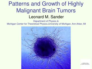
Patterns and Growth of Highly Malignant Brain Tumors Department of Physics & Michigan Center for Theoretical Physics,University of Michigan, Ann Arbor, MI Leonard M. Sander
Collaborators E. Khain1, A.M. Stein2, C. Schneider-Mizell Physics Department, University of Michigan M. O. Nowicki, E. A. Chiocca, S. Lawler Department of Neurological Surgery, The Ohio State University T. Demuth, M. E. Berens The Translational Genomics Research Institute, Phoenix, Arizona T. Deisboeck Complex Biosystems Modeling Laboratory, Harvard-MIT (HST); A. A. Martinos Center for Biomedical Imaging, Massachusetts General Hospital NIH grant R01 CA085139-01A2. Now at Oakland University, Michigan 2. Now at IMA, Minneapolis
Introduction to Malignant Brain Cancer • 18,000 people/year in the US are diagnosed with primary brain tumors. • 9,000 have glioblastoma multiforme (GBM), the most malignant form. • After diagnosis: • 50% of GBM patients die within 1 year. • 98% of GBM patients die within 5 years. • No significant advances in the last 30 years.
Why Glioblastoma has been Untreatable Pre-op. Post-op. 8 mo. • Surgery fails: • Cancer is highly invasive. • Some areas of the brain cannot be removed. • Chemotherapy and radiation fail: • Invasive cells proliferate slowly. • Blood-brain barrier blocks drug delivery.
Vocubulary: the word ‘model’ • A model for a physicist: • H = -ijSi•Sj • A model for a biologist:
Typical Invasion Models In vitro In vivo / In situ cell speed ~ 20 microns/hr
The 3d Tumor Spheroid Assay • Put a clump of cultured tumor cells (a tumor spheriod) in a gel. (We use collagen. • Spheriod grows. • Single cells invade. • A reasonable model for invasion in the brain. 3 mm Bright Field Image T. S. Deisboeck et. al. (2001) Pattern of self-organization in tumour systems: complex growth dynamics in a novel brain tumour spheroid model. Cell Prolif, 34, 115-134
Cell tracking A. M. Stein, D. A. Vader, L. M. Sander, and D. A. Weitz. Mathematical Modeling of Biological Systems, volume I. Birkhauser, 2006.
vr vθ Cells are Biased Random Walkers
Results of short-time tracking • Bias to move away from spheroid is clear, and decays in time. • Bias depends on cell line.
Longer-time behavior Day 1 Day3 Day5 Day7 U87dEGFR U87WT
Invasive cell motion has a random component and a directed component • Core radius expands at a “slow”, constant velocity. • Invasive cells are shed from the core surface • Invasive cells proliferate Rcore PDE Model Diffusion Directed Motility Cell Shedding Proliferation A. M. Stein, T. Demuth, D. Mobley, M. E. Berens, and L. M. Sander. A mathematical model of glioblastoma tumor spheroid invasion in a three-dimensional in vitro experiment. Biophys. J., 92:356–365, 2007.
Fit Model to Different Cells More malignant Less malignant
D v s g Sensitivity Analysis
What controls shed rate? • Cell cell adhesion is a good candidate. • Also, it probably controls clustering. WT dEGFR Cluster, possibly due to cell-cell adhesion. A secondary tumor?
Shed rate, clustering, and adhesion • Cells with large adhesion should have difficulty detaching from spheriod. • Clusters should result from adhesion. • Indirect measurement of adhesion through cell clustering. • Possible clinical significance: shed rate should correlate with invasiveness. • Can we use shed rate to guide surgery/ radiation, etc?
Simulations of clustering Phase separation and coarsening, q>qc 50 50 100 100 150 150 200 200 1 250 250 Phase separation 300 300 0.95 350 350 0.9 100 200 300 100 200 300 (A) (B) q, adhesion parameter 0.85 50 50 0.8 (C) (D) 100 100 150 150 0.75 No phase separation 200 200 0.7 250 250 0 0.1 0.2 300 300 c, average density 350 350 time No phase separation, q<qc 200 300
Experiments: Glioma cells on a surface Michal O. Nowicki, E. A. Chiocca, and Sean Lawler WT dEGFR No clustering Clustering Smaller cell-cell adhesion? (q<qc) Larger cell-cell adhesion? (q >qc)
Experiments II Michal O. Nowicki, E. A. Chiocca, and Sean Lawler dEGFR 1 day 3 days 5 days WT 3 days 1 day 5 days
Shed rate • We can measure the shed rate directly. • But, adhesion might also be important for secondary tumor formation.
Cause of Velocity Bias is Unknown • Chemotaxis • Nutrient gradients (glucose, O2) • Waste product gradients • Cell matrix interactions
Cell-Gel Interactions Two spheroids, 5mm apart D. Vader
A good model for cell-gel interactions requires a mechanical model for collagen
Single Cell in Collagen Vader and Weitz (Harvard)
Collagen is the primary animal structural protein. It is found in bone, cartilage, tendons, ECM, and jello. 1 nm ~100 nm
Collagen-I Gel 50 μm 1.5 mg/ml, from Vader and Weitz (Harvard)
Collagen Gel Physics Tension Test • Collagen is viscoelastic up to 10-15% strains. • Significant strain stiffening and plastic deformation occur at larger strains. • Many other biological gel networks have these properites, e.g. actin. • A micromechanical model is needed to understand strain stiffening and plasticity. Roeder et. al., 2002
Results on Actual Network Image Extended Branches Linked Branches
Tracking algorithm • Microscopy data to construct network.
Testing Algorithm withArtificial Networks • Seed space with fiber nucleation points • Chose random direction • Extend fibers along a persistent (lp) random walk • Create cross-link when two fibers are less than a fiber diameter (d) apart. • Stop extending fibers when the reach max length (L) cross-links
Testing Algorithm withArtificial Networks • All pixels within a radius (r) from the fiber backbone are set to one • To mimic confocal microscope, images are convolved with a gaussian point spread function, elongated in z
Extracting Artificial Networks True Network Black and White Image Convolved with PSF PSF + Noise
Extracting Artificial Networks True Network BW Image PSF PSF + Noise
Pinned Node Sliding Nodes Mechanical Modeling of Networks Impose Displacement elastic beams Minimize Energy
Minimize Total Energy Mechanical Modeling of Cross-links
0.1-50 Pa 0 0 0.05 Experimental ValidationSmall Strains Rigid Cross-Links Freely Rotating Cross-Links 1000-80000 Pa Kxlink 0 Pa
Estimation of Kxlink: Small Strainfull 3d network Kxlink (N-m)
Collagen Networks show Nonaffine Deformations 33 μm Free and Fixed cross-links More than 99% of energy in network is in bending
Strain Stiffening Model - 3d network projected to 2d Experiment 1.5 mg/ml 1.0 mg/ml 0.5 mg/ml 2 mg/ml
We seem to have forgotten about the cells Work in progress: • Treat cells as a force monopole or force dipole. • Look for characteristic length for deformation decay for single cell. • Model individual cell motility. • Look at fiber orientation decay for a spheroid. • Consider plastic deformations.
Summary • Lots of physics in glioma invasion. • Two processes: • Shedding of cells from tumor spheroids. • Depends on cell phenotype probably through cell-cell adhesion. • Motility. • Seems to depend on cell-environment interactions, at least in vitro. • First step in understanding cell-ECM interactions. • Mechanics of a collagen network.