Download
1 / 59
590 likes | 624 Vues
Three Types of Muscle Tissue. Skeletal muscle tissue: Attached to bones and skin Striated Voluntary (i.e., conscious control) Powerful Primary topic of this chapter. Three Types of Muscle Tissue. Cardiac muscle tissue: Only in the heart Striated Involuntary

E N D
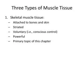
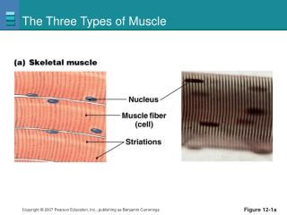
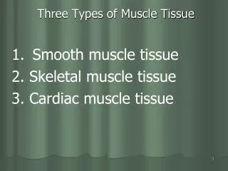
Three Types of Muscle Tissue • Skeletal muscle tissue: • Attached to bones and skin • Striated • Voluntary (i.e., conscious control) • Powerful • Primary topic of this chapter
Three Types of Muscle Tissue • Cardiac muscle tissue: • Only in the heart • Striated • Involuntary • More details in Chapter 18
Three Types of Muscle Tissue • Smooth muscle tissue: • In the walls of hollow organs, e.g., stomach, urinary bladder, and airways • Not striated • Involuntary
Special Characteristics of Muscle Tissue • Excitability (responsiveness or irritability): ability to receive and respond to stimuli • Contractility: ability to shorten when stimulated • Extensibility: ability to be stretched • Elasticity: ability to recoil to resting length
Muscle Functions • Movement of bones or fluids (e.g., blood) • Maintaining posture and body position • Stabilizing joints • Heat generation (especially skeletal muscle)
Skeletal Muscle • Each muscle is served by one artery, one nerve, and one or more veins
Skeletal Muscle • Connective tissue sheaths of skeletal muscle: • Epimysium: dense regular connective tissue surrounding entire muscle • Perimysium: fibrous connective tissue surrounding fascicles (groups of muscle fibers) • Endomysium: fine areolar connective tissue surrounding each muscle fiber
Epimysium Epimysium Bone Perimysium Endomysium Tendon Muscle fiber in middle of a fascicle (b) Blood vessel Fascicle (wrapped by perimysium) Endomysium (between individual muscle fibers) Perimysium Fascicle Muscle fiber (a) Figure 9.1
Skeletal Muscle: Attachments • Muscles attach: • Directly—epimysium of muscle is fused to the periosteum of bone or perichondrium of cartilage • Indirectly (more common) —connective tissue wrappings extend beyond the muscle as a ropelike tendon or sheetlike aponeurosis
Microscopic Anatomy of a Skeletal Muscle Fiber • Surrounded by sarcolemma (plasma membrane) • Long (huge) cylindrical cells (up to 30 cm!!!) • Multiple nuclei • Many mitochondria (Why So Many?) • Glycosomes (for glycogen storage) & myoglobin (for O2 storage) • Also contain myofibrils, sarcoplasmic reticulum, and T tubules
Myofibrils • Densely packed, rodlike elements (100’s to 1000’s per muscle fiber) • Makes up to 80% of muscle cell volume • Exhibit striations: perfectly aligned repeating series of dark A bands and light I bands
Sarcolemma Mitochondrion Myofibril Dark A band Light I band Nucleus (b) Diagram of part of a muscle fiber showing the myofibrils. Onemyofibril is extended afrom the cut end of the fiber.
Sarcomere • Smallest contractile unit (functional unit) of a muscle fiber • The region of a myofibril between two successive Z discs • Composed of thick and thin myofilaments made of contractile proteins responsible for muscle contraction
Features of a Sarcomere • Thick filaments: run the entire length of an A band • Thin filaments: run the length of the I band and partway into the A band • Z disc: coin-shaped sheet of proteins that anchors the thin filaments and connects myofibrils to one another • H zone: lighter mid-region on either side of the M line. Only seen in resting muscle fibers • M line: Found in the center of the H zone, it is a line of protein myomesin (M for middle)
Thin (actin) filament Z disc H zone Z disc Thick (myosin) filament I band A band Sarcomere I band M line (c) Small part of one myofibril enlarged to show the myofilaments responsible for the banding pattern. Each sarcomereextends from one Z disc to the next. Sarcomere Z disc Z disc M line Thin (actin) filament Elastic (titin) filaments Thick (myosin) filament (d) Enlargement of one sarcomere (sectioned lengthwise). Notice the myosin heads on the thick filaments. Figure 9.2c, d
Thick Filament(Myosin) • Composed of the protein myosin • Myosin tails contain: • 2 interwoven, heavy polypeptide chains • Myosin heads contain: • The “Business End” that act as cross bridges during contraction • Binding sites for actin of thin filaments • Binding sites for ATP • ATPase enzymes (split ATP to generate energy)
Thin Filament(Actin) • Composed mostly of protein actin • Bears active sites for the cross-bridges (heads of myosin) during contraction • Contains tropomyosin and troponin: regulatory proteins bound to actin • Elastic filament (made of titan protein) allows muscle to spring back into place
Longitudinal section of filaments within one sarcomere of a myofibril Thick filament Thin filament In the center of the sarcomere, the thick filaments lack myosin heads. Myosin heads are present only in areas of myosin-actin overlap. Thick filament Thin filament Each thick filament consists of many myosin molecules whose heads protrude at opposite ends of the filament. A thin filament consists of two strands of actin subunits twisted into a helix plus two types of regulatory proteins (troponin and tropomyosin). Portion of a thick filament Portion of a thin filament Myosin head Tropomyosin Troponin Actin Actin-binding sites Active sites for myosin attachment Tail Heads Actin subunits ATP- binding site Flexible hinge region Myosin molecule Actin subunits Figure 9.3
Sarcoplasmic Reticulum (SR) • Network of smooth endoplasmic reticulum surrounding each myofibril • Pairs of terminal cisternae (reservoirs) form perpendicular cross channels • Regulates calcium - stores and releases Ca+ for contraction (we’ll talk about Ca+ later)
T Tubules • Continuous with the sarcolemma • Penetrate the cell’s interior at each A band–I band junction • Associate with the paired terminal cisternae to form triads that encircle each sarcomere ***NOTE: A skeletal muscle is very long. T-tubules allow the electrical stimulus and ECF to come in contact with deep regions which makes muscle reaction occur quicker
Part of a skeletal muscle fiber (cell) I band A band I band Z disc H zone Z disc Myofibril M line Sarcolemma Triad: T tubule • • Terminal cisternae of the SR (2) Sarcolemma Tubules of the SR Myofibrils Mitochondria Figure 9.5
CHECK POINT!!!!! 1.) Which myofilaments have heads that form cross-bridges that are important during contraction? Thick filaments 2.) What surrounds the myofibril and regulates Ca+ needed for contraction? Sarcoplasmic Reticulum
You have 7 minutes from the time the bell rings…. If you don’t turn it in on time, you don’t get the credit! Briefly explain how actin and myosin work together in the sliding filament model Explain the major role of the sarcoplasmic reticulum (SR), especially the terminal cisternae. What key substance provides the final “go” signal for contraction? Hint: pg 282 What structure works close with the SR and forms a triad relationship?
Contraction • The generation of force • Shortening occurs when tension from the cross bridges on the thin filaments pulls the thin filament toward the M line ultimately contracting or shortening the muscle fiber
Sliding Filament Model of Contraction • In the relaxed state, thin and thick filaments only overlap slightly • During contraction, myosin heads bind to actin, detach, and bind again, to propel the thin filaments toward the M line • As H zones shorten and disappear, sarcomeres and muscle cells shorten, thus, the whole muscle shortens
Sarcomere Contraction Animation Z Z H A I I 1 Fully relaxed sarcomere of a muscle fiber Z Z I A I 2 Fully contracted sarcomere of a muscle fiber Figure 9.6
So now we know how a muscle fiber contracts…but what causes it to contract? • Activation: neural stimulation at aneuromuscular junction • Excitation-contraction coupling: • Creation and increase of an action potential (electrical current) along the sarcolemma • Final trigger: a brief rise in intracellular Ca+ levels
Step One- The Activation Step: Takes place at the Neuromuscular Junction • Skeletal muscles are stimulated by somatic motor neurons • Axons of motor neurons travel from the central nervous system (brain or spinal cord) via nerves to skeletal muscles • Each axon forms several branches as it enters a muscle • Each axon ending forms a neuromuscular junction with a single muscle fiber
The axon of each motor neuron divides profusely and forms a neuromuscular junction at each muscle fiber
Myelinated axon of motor neuron Action potential (AP) Axon terminal of neuromuscular junction Nucleus Sarcolemma of the muscle fiber 1 Action potential arrives at axon terminal of motor neuron. Ca2+ Synaptic vesicle containing ACh Ca2+ 2 Voltage-gated Ca2+ channels open and Ca2+ enters the axon terminal. Mitochondrion Synaptic cleft Axon terminal of motor neuron Fusing synaptic vesicles Figure 9.8 Figure 9.8
Neuromuscular Junction • Situated midway along the length of a muscle fiber • Axon terminal and muscle fiber are separated by a gel-filled space called the synaptic cleft • Synaptic vesicles within the axon terminal contain the neurotransmitter acetylcholine (ACh) • Junctional folds of the sarcolemma contain ACh receptors
Events at the Neuromuscular Junction • Nerve impulse arrives at axon terminal • Voltage sensitive Calcium channels open and release Calcium into the axon terminal • Due to increased Calcium levels, ACh is released and binds with receptors on the sarcolemma which triggers electrical events • These electrical events lead to the creation of an action potential (electrical current) which spreads down the sarcolemma
Destruction of Acetylcholine • ACh effects are quickly stopped by the enzyme acetylcholinesterase which is located in the synaptic cleft • Prevents continued muscle fiber contraction by breaking down Ach into basic “non-stimulating” components
Myelinated axon of motor neuron Action potential (AP) Axon terminal of neuromuscular junction Nucleus Sarcolemma of the muscle fiber 1 Action potential arrives at axon terminal of motor neuron. Ca2+ Synaptic vesicle containing ACh Ca2+ 2 Voltage-gated Ca2+ channels open and Ca2+ enters the axon terminal. Mitochondrion Synaptic cleft Axon terminal of motor neuron 3 Ca2+ entry causes some synaptic vesicles to release their contents (acetylcholine) by exocytosis. Fusing synaptic vesicles Junctional folds of sarcolemma ACh 4 Acetylcholine, a neurotransmitter, diffuses across the synaptic cleft and binds to receptors in the sarcolemma. Sarcoplasm of muscle fiber Postsynaptic membrane ion channel opens; ions pass. 5 ACh binding opens ion channels that allow simultaneous passage of Na+ into the muscle fiber and K+ out of the muscle fiber. K+ Na+ Degraded ACh 6 ACh effects are terminated by its enzymatic breakdown in the synaptic cleft by acetylcholinesterase. Ach– Postsynaptic membrane ion channel closed; ions cannot pass. Na+ Acetyl- cholinesterase K+ Figure 9.8
Axon terminal Open Na+ Channel Closed K+ Channel Synaptic cleft Na+ ACh K+ Na+ K+ + + + + ACh + + + + + + Action potential n + + o i Na+ K+ t a 2 Generation and propagation of the action potential (AP) z i r a l o p e d f o e v Closed Na+ Channel Open K+ Channel a W 1 Local depolarization: generation of the end plate potential on the sarcolemma Na+ K+ 3 Repolarization Sarcoplasm of muscle fiber Figure 9.9
Axon terminal Open Na+ Channel Closed K+ Channel Na+ Synaptic cleft ACh K+ Na+ K+ + + + + ACh + + + + + + Action potential n + + o i t Na+ K+ a z 2 i r Generation and propagation of the action potential (AP) a l o p e d f o e v a W 1 1 Local depolarization: generation of the end plate potential on the sarcolemma Sarcoplasm of muscle fiber Figure 9.9, step 2
Excitation-Contraction (E-C) Coupling • Sequence of events by which transmission of an AP along the sarcolemma leads to sliding of the myofilaments • Occurs during latent period: • Time between AP initiation and the beginning of contraction
Events of Excitation-Contraction (E-C) Coupling • AP is spread along sarcolemma to the T tubules • Voltage-sensitive proteins stimulate the SR to release Ca+ • Ca+ is necessary for contraction
Setting the stage Axon terminal of motor neuron Action potential is generated Synaptic cleft ACh Sarcolemma Terminal cisterna of SR Ca2+ Muscle fiber Triad One sarcomere Figure 9.11, step 1
Steps in E-C Coupling: Sarcolemma Voltage-sensitive tubule protein T tubule Action potential is propagated along the sarcolemma and down the T tubules. 1 Ca2+ release channel 2 Calcium ions are released. Terminal cisterna of SR Ca2+ Actin Tropomyosin blocking active sites Troponin Ca2+ Myosin 3 Calcium binds to troponin and removes the blocking action of tropomyosin. Active sites exposed and ready for myosin binding 4 Contraction begins Myosin cross bridge The aftermath Figure 9.11, step 2
1 Action potential is propagated along the sarcolemma and down the T tubules. Steps in E-C Coupling: Sarcolemma Voltage-sensitive tubule protein T tubule Ca2+ release channel Terminal cisterna of SR Ca2+ Figure 9.11, step 3
1 Action potential is propagated along the sarcolemma and down the T tubules. Steps in E-C Coupling: Sarcolemma Voltage-sensitive tubule protein T tubule Ca2+ release channel 2 Calcium ions are released. Terminal cisterna of SR Ca2+ Figure 9.11, step 4
Actin Troponin Tropomyosin blocking active sites Ca2+ Myosin The aftermath Figure 9.11, step 5
Actin Troponin Tropomyosin blocking active sites Ca2+ Myosin 3 Calcium binds to troponin and removes the blocking action of tropomyosin. Active sites exposed and ready for myosin binding The aftermath Figure 9.11, step 6
Actin Troponin Tropomyosin blocking active sites Ca2+ Myosin 3 Calcium binds to troponin and removes the blocking action of tropomyosin. Active sites exposed and ready for myosin binding Contraction begins 4 Myosin cross bridge The aftermath Figure 9.11, step 7
Steps in E-C Coupling: Sarcolemma Voltage-sensitive tubule protein T tubule Action potential is propagated along the sarcolemma and down the T tubules. 1 Ca2+ release channel 2 Calcium ions are released. Terminal cisterna of SR Ca2+ Actin Tropomyosin blocking active sites Troponin Ca2+ Myosin 3 Calcium binds to troponin and removes the blocking action of tropomyosin. Active sites exposed and ready for myosin binding 4 Contraction begins Myosin cross bridge The aftermath Figure 9.11, step 8
Role of Calcium (Ca2+) in Contraction • At low intracellular Ca2+ concentration: • Tropomyosin blocks the active sites on actin • Myosin heads cannot attach to actin • Muscle fiber relaxes
Role of Calcium (Ca2+) in Contraction • At higher intracellular Ca+ concentrations: • Ca+ binds to troponin • Troponin changes shape and moves tropomyosin away from active sites • Events of the cross bridge cycle occur • When nervous stimulation stops, Ca+ is pumped back into the SR and contraction ends