Download
1 / 49
1.14k likes | 2.33k Vues
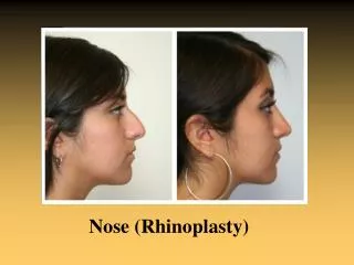
Anatomical approach to rhinoplasty. Herve LeBoeuf, MD Karen Calhoun, MD. Which Incision ??. Transfixion/Hemitransfixion. Caudal septum, medial crura, nasal spine Just caudal to septum Follows medial crura to flared ends Extend to floor for tip projection access Hemi- is unilateral only

E N D
Anatomical approach to rhinoplasty Herve LeBoeuf, MD Karen Calhoun, MD
Transfixion/Hemitransfixion • Caudal septum, medial crura, nasal spine • Just caudal to septum • Follows medial crura to flared ends • Extend to floor for tip projection access • Hemi- is unilateral only • Avoids disruption of tip support • Poorer access, ? Asymmetric healing
Intercartilaginous Incision • Access to the tip and mid-nose • Incision intranasal, between the ULC/LLC • Begin medially as transfixion extension • Continue entire length of LLC • Avoid transecting the lateral end of the LLC
Intracartilaginous Incision • Access to the tip and mid-nose • Incise through vestibular mucosa +/- lower lateral cartilage • Similar to intercartilaginous, but 3-5mm caudal to the cephalic end of LLC • This is caudal to the nasal valve • Decreases risk of nasal obstruction (avoids scar contracture of the valve)
Rim and Marginal Incisions • Made parallel to the caudal borders of LLC (cephalic border of nasal vibrissae) • Endonasal approach • More access to modify LLC • Combined with intercartilaginous incision to create pedicled or bipedicled flap of cartilage and mucoperichondrium • Always used in external approach • Extend to lateral end of LLC • In continuity with the transfixion incision
Transcollumellar • External approach • Crosses collumella just above flared ends of the medial crura • If too close to the lip, “dip” deformity • No cartilage support to counteract tension generated by the healing skin • Notching at the midline – “aggie mark”, Improved scar camouflage
Lateral Osteotomy • Access for the osteotomy • Short stab incisions just anterior to anterior attachment of the anterior turbinate • Directed deep and laterally toward the bony piriform aperture • +/- subperiosteal tunnels for osteotome
Open verses Closed ??? • Open • Much better exposure of structures • More accurate placement of grafts • More accurate structural diagnosis • Teaching value • Closed • Possibly faster than open • No external scar • Avoids tip edema • No loss of tip support
Nasal Tip – Lower Lateral Cartilage • Paired to form arch supporting lobule/nostrils • Divided into medial and lateral crura • Lateral crura • Flare posterosuperiorly away from rim • Tip defining point – junction between central and lateral crura • Medial crura • Joined by ligamentous tissue in columella • Sagittal orientation with caudal flaring • Collumellar double break: medial crus bends posteriorly at superior extent, marks beginning of the central crus
Nasal Tip • Dome: formed by the junction of the medial and lateral crura • Two point tip: aesthetically pleasing • Tent deformity: Single point tip • Overtight suture or poorly placed tip graft • Sesamoid Cartilage • Accessory cartilage between lateral crura and piriform aperture • Cephalic border of the lower lateral cartilage forms hinge with upper lateral cartilage
Tip Support • Anderson: nasal tip similar to a Tripod • Conjoined medial crura and two lateral crura represent the three legs of the tripod • Major support • Size, shape, resilience of medial and lateral crura • Fibrous attachment of the medial crura feet to the caudal septum • Fibrous attachment of the caudal margin of the ULC to the cephalic margin of the LLC
Tip Support • Minor Support • Ligamentous sling between the alar cartilages • Cartilaginous septal dorsum • Sesamoid complex – extending the support of the lateral crura to the piriform aperture • Attachment of the alar cartilages to overlying skin and musculature • Nasal spine • Membranous septum
Upper Lateral Cartilages • Triangular, base at septum/ apex at pyriform • Cephalic attachment to nasal bones • Nasal bones overlap ULC 1cm • Held in place with ligamentous fibers • Attached to septum medially, which broadens to form a platform for the cartilages • Intranasal valve: junction of ULC with septum • Ligaments connect with pyriform laterally to hold valve open, may be damaged during rhinoplasty and result in nasal obstruction
The Wide or Bulbous Tip • Excess amount and/or convex curvature of the cephalad alar lateral crus • Lateral alar convexities causing a trapezoid appearance from the basilar view • Increased interdomal distance • Poor dome definition – often due to excessively obtuse angle between the medial and lateral crus
Excessive Cephalad Alar Cartilage • Incise the cartilage • Incise and morselize the cephalad cartilage • Excise the cephalad cartilage
Goal: Unified Symmetric Tip • Med crura fixation stitch • Stabilizes crura during strut placement • Collumellar strut • Maintains columellar shape • Flare Control Sutures • Narrow width of columella by decreasing crural flare after strut
Goal: Correct Lateral Alar Convexity • Lateral crura spanning suture • Dome spanning suture
Tip Projection • posterior to anterior distance that the tip defining point extends from the facial plane at the alar crease
Tip Rotation • Movement of the tip along a circular arc consisting of a radius centered at the nasolabial angle that extends to the tip defining point
Increasing Projection • Columellar strut, +/- flare control suture • “Projection Control Suture”….advancement • Intradomal / interdomal suture
Increasing Projection • Trim protruding caudal septum, if any • Add tip graft if the infratip lobule becomes overshortened
Decreasing Projection • Collumellar Strut, Flare sutures if needed • Projection control sutures….recessive • If lateral alar convexity, correct with interdomal suture
Decreasing Projection • Intradomal stitch, if needed to correct widened domes • May need to transect lateral crura • May need to address medial crural or alar flaring
Some Tip Rotation Maneuvers • Cephalic trim of LLC • Weakens tip support by dividing ligaments between ULC and LLC, may cause bossae • Excise triangle of cartilage from mid LLC • Lateral Crural Steal • Illusion of rotation • Tip grafts • Lowering of dorsum