Deep Vein Thrombosis (DVT)
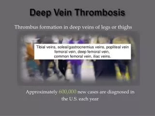
Deep Vein Thrombosis (DVT). Formation of a clot within a vein Occurs when there is i ) venous stasis ii)Vessel injury ii)hyper coaguability. References.

Deep Vein Thrombosis (DVT)
E N D
Presentation Transcript
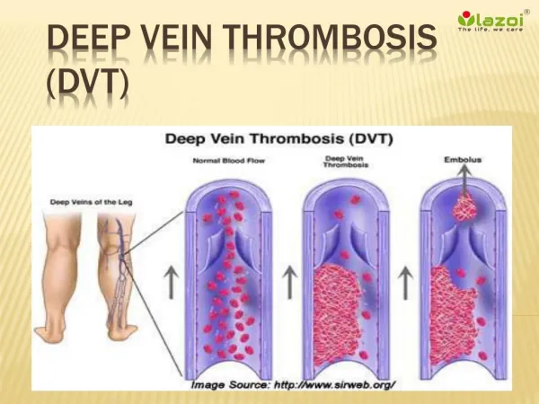
Deep Vein Thrombosis (DVT) • Formation of a clot within a vein • Occurs when there is i) venous stasis • ii)Vessel injury • ii)hyper coaguability
References • Myer, Kenneth A. and Amy Clough. Making Sense Of Vascular Ultrasound: A Hands-on Guide. London: Arnold, 2004 • Allan, Paul L.P. Clinical Doppler Ultrasound [Oxford]: Churchill Livingstone/ Elsevier, 2006 • 2) http://sonoworld.com/Client/Lectures/LectureDetails.aspx?Id=973&Sequence=1 • Online evidence accessed 1/10/13 • Bruno, G. L. 2013. Looking beyond the routine DVT scan. Soundeffects 1
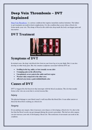
Predisposing factors • VENOUS STASIS • Prolonged periods of inactivity • Limb immobilisation due to fracture • Post surgery immobilisation • Cancer • VESSEL INJURY • Causing non functioning of valves • HYPERCOAGUABILITY • Congenital thropmophilia
Presentation • Usually asymptomatic • Calf swelling • Pain, worse when flexing the foot • redness
Ultrasound appearances Normal veins Vein with thrombus Static echoes within the lumen Incomplete or absent colour within lumen on colour doppler Partial or non compressibility Loss of spontaneous phasic flow with respiration Increased flow collateral vessels • Anechoic lumen • Colour doppler fills the whole lumen • Compressibility • Phasic flow with respiration • Very few or no collateral channels
When there is a thrombus • Image of non compressible vein • Its location on the leg in relation to a landmark. ( Groin crease, knee crease?) • Is it in a deep or superficial vein? • If in superficial vein, its distance from the junction with the deep vein
Colour Flow Doppler Showing DVT • Absence of colour flow in the peroneal branches
The Report • Size and position of thrombus • Distance of thrombus from either groin or knee • Age thrombus