DEEP VEIN THROMBOSIS
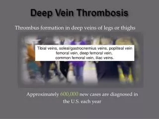
DEEP VEIN THROMBOSIS. Definitions. Deep-vein thrombosis (also known as deep-venous thrombosis or DVT) is the formation of a blood clot ("thrombus") in a deep vein. Pulmonary embolism (PE) is a condition that occurs when an artery in your lung becomes blocked. Thrombosis. Virchow’s Triad.

DEEP VEIN THROMBOSIS
E N D
Presentation Transcript
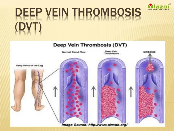
Definitions • Deep-vein thrombosis (also known as deep-venous thrombosis or DVT) is the formation of a blood clot ("thrombus") in a deep vein. • Pulmonary embolism (PE) is a condition that occurs when an artery in your lung becomes blocked.
Signs & Symptoms • Pain in the leg • Tenderness in the calf (one of the most important signs) • Leg tenderness • Swelling of the leg • Increased warmth of the leg • Redness in the leg • Bluish skin discoloration
DIAGNOSIS OF DVT • Clinical examination • Investigations
CLINICAL EXAMINATION • Homan’s sign • Moses’ sign • Phlegmasia alba dolens (white leg) • Phlegmasia cerulea dolens (blue leg)
Diagnosis of DVT Duplex Ultra sonography -Projected sound waves bounce off structures in the leg and create images that reveal abnormalities. The addition of color Doppler imaging improves accuracy. Venography – An x-ray of leg and pelvis will show the calf and thigh veins and reveal any blockages.
Diagnosis of DVT MRI - particularly effective in diagnosing DVT in the pelvis, and as effective as venography in diagnosing DVT in the thigh. Cuff-impedance plethysmography -uses blood pressure checks at different places in the leg to identify possible blockages.
Potential Complications • Pulmonary emboli- most serious complication of DVT • Chronic venous insufficiency - Long-term DVT can degenerate the venous valves. • Post- phlebotic syndrome - long-term complication of DVT which occurs due to damage and scarring to the veins and is characterized by swelling, discomfort and skin pigmentation in the affected area.
DVT Risk Factors Risk Factor= 1 Point Risk Factor= 2 Points • Age 41-60 years • Bedrest • COPD • CHF (<1 month) • Acute MI • Pneumonia (< I month) • Sepsis (<month) • Inflammatory Bowel Disease • Minor surgery planned • Hx of prior major surgery • Obesity (BMI>25) • Swollen legs • Pregnancy • Oral Contraceptives • Age 60-74 years • Athroscopic surgery • Laparoscopic surgey • Malignancy (present or history) • Major surgery (>45 Minutes) • Patient confined to bed (>72 hours) • Immobilizing plaster cast(<1month) • Central Venous Access • Infection • Nephrotic Syndrome
DVT Risk FactorsRisk Factor= 3 Point Risk Factor =5 Points • Age >75 years • Major surgery(>3 hours) • Hx of DVT/PE • Family hx of Thrombosis • Heparin-induced thrombocytopenia • Thrombophilia • Hx of clotting disorder • Elective major lower extremity arthroplasty • Hip, pelvis, or leg fracture(<1 month) • Stroke(< 1 month) • Acute spinal cord injury (paralysis<1 month)
RISK LEVEL FOR DVT….. • Low Risk- DVT score 1 • Moderate Risk- DVT score 2 • High Risk- DVT score 3-4 • Highest Risk- DVT score 5 or more
Prevention DVT/PE • Pharmacologic • Non-pharmacologic
Prevention: Non-pharmacologic Mechanical methods: • Used in combination with drug therapy or as monotherapy in those with lower risk or contraindications to anticoagulation prophylaxis • Includes aggressive mobilization, foot pumps, intermittent pneumatic leg pumps, graduated compression stockings • These methods MUST be used for most of the day (>21 hours) to be effective
Prevention: Pharmacologic • Unfractionated Heparin • Low Molecular Weight Heparin (LMWH) • daltaparin (Fragmin), enoxaparin (Lovenox) • Factor Xa Inhibitor • fondaparinux (Arixtra) • Warfarin (Coumadin)
Contraindications to Pharmacological Prophylaxis • Active GI bleed • Recent hemorrhagic stroke or hemorrhage • Previous hypersensitivity or significant drug-specific ADR • Platelet count <100 X 109/L (varies) • Neurosurgical Procedure within last 30 days • Varies between institutions, physicians, and particular patient risk/benefit considerations
Unfractionated Heparin • Indications: Prophylaxis and treatment of thromboembolic disorders • Prophylactic Dose: • 5,000 Units SQ q8hrs • Monitoring: PTT, Plt, Hgb, Hct, bleeding
Unfractionated Heparin • Complications: hemorrhage (most common), thrombocytopenia, hyper-sensitivity • Advantages: Cost, DVT prophylaxis of choice in pregnancy
Warfarin (Coumadin) • Indication: Treatment of venous thrombosis, pulmonary embolis, and thromboembolic disorder • Not generally used for DVT/PE prophylaxis (except long term prevention, such as patients with valve replacements) due to delayed onset of action and higher risk of bleeding complications • Dose: • Typical starting dose is 2.5 to 5 mg • Use “bridge therapy” (LMWH or heparin + warfarin) when immediate anticoagulation is warranted
Bridge Therapy • The anticoagulant effects of warfarin (Coumadin) are not immediate • It takes several days (~ 5 days) to a week to deplete existing Vitamin K dependent clotting factors in the body • During this lag time, the patient must be protected against DVT/PE with Heparin, enoxaparin (Lovenox), daltaparin (Fragmin) or fondaparinux (Arixtra) until the therapeutic INR is achieved
Bridge Therapy • Concurrent therapy with warfarin (Coumadin) MUST be continued for a minimum of 5 days, AND 2 consecutive INR values must be seen before the discontinuation of LMWH or heparin may occur