Advanced Microstructural Characterization of Dimpled Laminated Metals
This research explores the innovative technique of dimpling for microstructural characterization of laminated metals, particularly steel and brass. By employing advanced sample preparation methods, including ion milling and the use of specialized grinding wheels, we aim to achieve optimal specimen thickness and surface quality for electron microscopy. The study assesses the mechanical properties and microstructural features of these multilayer laminates, laying groundwork for future investigations and potential publications in material sciences.

Advanced Microstructural Characterization of Dimpled Laminated Metals
E N D
Presentation Transcript
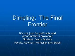
Dimpling: The Final Frontier It’s not just for golf balls and grandmothers anymore! Student: Jason Burbey Faculty Advisor: Professor Eric Stach
Outline • Background • Objective • Sample Preparation/Characterization • Sample Prep. Instruments • Work Completed • Future Work • Acknowledgements • Questions
Background • Professor Ravi Chandran • University of Utah • Laminated Steel and Brass • Repeated cold rolling • Continuation of previous study • Evaluated Mechanical Properties • Inverse relationship between yield strength and bilayer thickness • Yield strengths as high as 850 MPa • Why? • Microstructural characterization needed Ravichandran, K. “Nanoscale Steel-Brass Multilayer Laminates Made By Cold Rolling: Microstructure and Tensile Properties.” Scripta Materialia Vol 42: 949,951
Objective • Microstructural Characterization of laminates • Bright Field Images in TEM • Features of interest: • Bilayer thickness • Interlamellar structure • Dislocation density and distribution
Sample Preparation/Characterization • Dimple specimens to 10-15um • Ion Mill to produce very small hole in specimen • Electron Transparent • Take bright-field images of specimen using TEM
The Dimpler • Model 2000 Sample Prep System • Diamond Slurries and pastes • 6,3,1, .25 micron
The Ion Mill • Gatan Model 600 Duo-Mill • Ar+ ions directed at specimen from both sides at a set angle • Fairly time consuming
Transmission Electron Microscope • JEOL Model 2000FX • Uses an Ultra High Vacuum (UHV) • 10^-9 -10^-11 atm • Capable of high resolution images at high magnifications • 1,000,000X or higher • Resolution of < 3Å
Work completed • Dimpled metallic laminates • SS grinding wheel • Ion Mill attempt • Micarta grinding Wheel • Dimpled Ta/Si specimens • Dimpled InAs/GaP specimens
Metallic Laminates: SS grinding Wheel • Thicknesses varied from 90-290um • Used 3um slurry and dimpled to 20um • Not all specimens were 20um • Used optical microscope to check thickness • Re-dimpling difficult • Specimens 10-15um achieved • Ion Mill trials • Inefficient • Start Over! • Made more specimens • New dimpling progression • Used different wheel
Dimpling Laminates: Micarta Grinding Wheel • Micarta Wheel is flatter and thicker than the SS grinding wheel • Dimple the whole specimen surface • Used 6um slurry • Sped up dimpling progress • Much smoother surface • Very important for Ion Milling • Successful specimens created • Stopped after 1um slurry • More scratches observed with .25um paste
Ta/Si Specimens • Checking phase transformation of Ta • Blue side = Si • Reflective = Ta • Use diamond scribe to cleave specimens
Dimple on Si side SS grinding wheel Specimens thicker than laminates 470-520um Same progression as laminates Same force exerted by grinding arm Sample Prep Problem • Destruction of specimens • Force too much • Si is fragile! • Would crack at approx. 20um
Solution for Ta/Si specimens • Use less force! • Progressively decrease force • Before switching slurry/paste • 3 specimens from each bulk sample ready for Ion Milling
In/As specimens • Thicknesses between 310-320um • Dimpled on GaP side • Used SS grinding wheel • Same dimpling progression as Ta/Si • Like butter! • It smells… • 3 specimens ready for Ion Milling
Future Work • Ion Mill all of the different specimens • Characterize specimens in TEM with help from Professor Stach • Publish characterization results
Acknowledgements • Dr. King • Professor Stach • Cheryl Waller • Vijay Rawat • Rocco Cerchiara from Fischione • Jan Eberle • MSE Dept.