Download
1 / 37
450 likes | 967 Vues
Metabolic and endocrine disorders in pet animals. N.K. Rakha 1 , V. K. Jain 2 and Rajesh Khurana 1 1 Teaching Veterinary Clinical Service Complex; 2 Veterinary Clinical Medicine CCS Haryana Agricultural University, Hisar. Metabolic and endocrine disorders. Rickets Osteomalacia

E N D
Metabolic and endocrine disorders in pet animals N.K. Rakha1, V. K. Jain2 and Rajesh Khurana1 1Teaching Veterinary Clinical Service Complex;2Veterinary Clinical Medicine CCS Haryana Agricultural University, Hisar
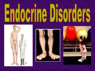
Metabolic and endocrine disorders • Rickets • Osteomalacia • Osteoporosis • Hypo-phosphataemia • Hyper-phosphataemia • Diabetes mellitus • Diabetes insipidus • Nutritional secondary hyper-parathyroidism • Renal secondary hyper-parathyroidism
1. Rickets Vitamin D is required for orderly growth & mineralization of epiphysis (growth plate). Young animals fed deficient diets or housed indoors without exposure to UV radiation develop rickets.
Pathogenesis There may be a decrease in serum Ca & P levels due to impaired intestinal absorption. Mineralization of cartilaginous matrix fails to occur and physis become irregularly thickened.
P deficiency also will result in rickets because of the failure to maintain an adequate ion product of serum Ca and P at the zones of mineralization in bone. • P deficiency will not results in hypocalcaemia & serum levels will be normal or increased.
Clinical findings • Long bones of the legs get bent. • There is beading over ribs and long bones • In severe cases there is lameness associated with enlargement of the ends of fast growing bones and deformities of the weight bearing long bones.
Clinical diagnosis • Lameness and enlargement of the joints is more commonly due to chronic polyarthritis and in young large dogs due to hypertrophic osteodystrophy. • Radiograph of rachitic bones show wide growth plates and demineralization.
Treatment Administration of • Vitamin D, • Ca & • P leads to re-establishment of normal calcification in osteoid on bone surfaces and physes.
2. Osteomalacia It literally means, “softening of bones”. Characterized by defective mineralization of osteoid (bone matrix) in mature bone due to deficiency of Vit D or P. Much less inhibition of bone matrix formation compared to impairment of matrix mineralization. Adequate local conc. of P & Ca is necessary for proper deposition of minerals at mineralization front.
Pathogenesis • P deficiency inhibits normal osteoblast function and reduces the Ca – P ion product. • Vitamin D stimulates Ca and P absorption from the intestine and help in normal osteoblast function.
Diagnosis • Diagnosis of osteomalacia required interpretation of bone pain, serum Ca, P & Vit D conc. • Radiography and bone biopsy will also help in diagnosis. • Osteomalacia may also occur during fat malabsorption syndrome due to inhibition of absorption of Ca, P & Vit D.
3. Osteoporosis It is defined as a reduction in bone density that predisposes bone to fracture (however, bone is structurally normal). It may occur as a result of malnutrition, deficiency of Ca or P, during protein wasting diseases (e.g. parasitism), local or generalized skeletal disuse or as an aging change.
4. Hypo-phosphataemia [ Hypo-phosphataemia can be termed when serum P conc. < 3 mg/dl in dogs and cats.] Etiology: It may result from • Decreased absorption of phosphate from the intestines, • Increased urinary phosphate excretion e.g. hyper-parathyroidism or • Shift of phosphate from the extracellular to intracellular compartment e.g. after the treatment of Diabetic keto acidosis.
Clinical features • In most of cases it affects haematological & neuromuscular system in dogs & cats • Hemolytic anaemia is the most common sequel of the hypo-phosphataemia and may be life threatening in the absence of treatment • Other signs observed may be weakness, ataxia and seizures.
Treatment Remove the underlying cause. • Phosphate may be administered orally (Potassium or Sodium salt, according to the situation. • In severe cases i.v. in NSS. (0.01 -0.03 mmol/kg/hr). • Avoid hyperphosphataemia; therefore frequent monitoring of phosphate is advisable.
5. Hyperphosphataemia When serum phosphate (PO4) conc. is greater than 6.5. mg/dl in adult dogs. Dogs and cats under 6 months of age normally have higher serum phosphate concentration than adults.
Etiology • It can result from increased intestinal phosphate absorption, decreased phosphate excretion in urine, or shifting of phosphate from intra-cellular to extra-cellular compartment. • Most common causes in dogs/cats are renal failure, Vit D toxicosis (i.e. cholecalciferol / rodenticide toxicosis) and hypophosphataemia.
Clinical features • It reflects some other underlying disease. • By itself, it usually doesn’t cause clinical signs. • An acute increase in serum phosphate level may cause hypo-calcaemia. • Sustained hyperphosphataemia can cause 20 hyper-parathyroidism, fibrous osteo-dystrophy and metastatic calcification in exosteoseous sites. • Sustained hyperphosphataemia is usually caused by renal insufficiency or hypo-parathyroidism.
Treatment: • Treat underlying disease (renal failures) • Give protein restricted diets • Oral administration of phosphate chelating gels. (Aluminium preparations)
6. Diabetes mellitus Insulin dependent Diabetes mellitus is characterized by • Hypo-insulinaemia, • Poor glycaemic control in response to diet or treatment with oral hypo-glycaemic drugs or both and • Need of exogenous insulin to maintain the glycaemic control.
Etiology Causes are poorly characterized & multi-factorial e.g. • Genetic pre-disposition, • Infection, • Insulin antagonistic diseases and drugs, • Obesity, • Immune mediated pancreatitis. This results in impaired transport of circulating glucose into most of cells, accelerated hepatic gluconeogenesis and glycogenolysis.
In turn, this leads to hyperglycaemia, glycosuria, polyuria, polydipsia, polyphagia, weight loss, ketoacidosis (over production of ketone bodies to compensate for the underutilization of blood glucose). • A transient or reversible form of IDDM may be seen in bitches in diestrus due to insulin antagonism.
Clinical features • Peak prevalence at 7-9 yr of age (range 4-14 yrs). • Juvenile diabetes (<1 year) is uncommon. • Females suffer twice than males. • Breed disposition is observed • Pulik, Cairn Terriers, Miniature Pinschers, poodles, Dachshunds, Miniature schnanzers and beagles are more affected • Cocker spaniel, German Shephard, Collies, Pekingese, Rotweilers, and Boxers are relatively less affected.
History: • Hyperglycaemia results in glycosuria, which may be reflected as polyuria, polydipsia, polyphagia and weight loss. • There is frequent need to change the litter. • There is development of sudden blindness or rear limb weakness, plantigrade posture (hocks touch the ground when cat walks, differentiate from arthritis). • Later on, animal may develop ketonaemia, metabolic acidosis or diabetic ketoacidosis
Clinical features • Animal may be obese or in good physical condition. • In later stages, wt. loss may be there or it may be due to some other concurrent disease like pancreatic exocrine insufficiency, hyper thyroidism. • Hepatic lipidosis results in hepathomegaly. • Fasting hyperglycaemia (> 130 mg/dl) & glycosuria are important laboratory findings. Cataract is also a coomon clinical finding in diabetic dogs.
Treatment : Important goal is to maintain near normoglycaemia with help of • Medicines (sulfonyl ureas, glipizide, biguanides), • Insulin, • Diet & • Exercise.
7. Diabetes insipidus Arginine vasopressin (AVP) has an important role in water balance, renal water resorption, urine production and concentration. It is stored and secreted from the posterior pituitary gland and acts on distal tubular and collecting duct cells of kidney. Diabetes inspidus (DI) is caused as a result of defective synthesis or secretion of AVP (Central DI) or due to inability of the renal tubules to respond to AVP (Nephrogenic DI). There is no age, breed or sex related predilection for DI.
Etiology • The causes of CDI are trauma, idiopathic, neoplasia, pituitary malformation, cyst, inflammation etc. • While NDI may be primary idiopathic, primary familial (huskies) or secondary acquired renal insufficiency or failure, hyperadrenocorticism, hypoadrenocorticism, hepatic insufficiency, pyometra, hypercalcaemia, hypokalaemia, post obstructive diuresis, normo-glycemic glucosuria, hyper-thyroidism.
Clinical signs • Polyuria and polydipsia are the hallmark signs of diabetes inspidus. • Nervous signs may be seen in animals in which hypothalamic or pituitary tumor may be growing in the head. • The urine sp. gravity is 1.005 (Range 1.001 – 1.012)
Treatment: Synthetic analog of vaso-pressin as nasal spray or eye drops is reported to be very effective (1-4 drops o.d. or b.i.d.) or s.c. administration may be practiced (0.5 – 2 µg, o.d. or b.i.d.)
8. Nutritional secondary hyper-parathyroidism It is nutritional/ metabolic disorder caused by elevation of parathyroid hormone (PTH), usually secondary to poor nutrition. All meat or all grain diet is rich in P which is responsible for this disorder. Low calcium and Vitamin D and malabsorption also cause hypocalcaemia.
Increased PTH causes bone resorption, & Ca, thus, mobilized is transferred to extra-cellular fluid. Depletion of bone osteoid leads to deformities, fractures and loss of structural support.
Clinical Picture: • Young animals suffer lameness/ bone deformities • Spontaneous fractures of long bones or vertebrae occur many times. • Increase in serum phosphate, alkaline phophatase & PTH is there but Ca may be normal or decreased. • Radiography show systemic bone resorption/ fracture
Treatment: • Ca supplementation & dietary correction is must. • Restrict the movements to prevent fractures. • External splint should be applied.
9. Renal Secondary hyper-parathyroidism • This is caused by increased PTH levels associated with congenital/ acquired renal insufficiency. • Due to impaired P excretion and diminished conversion of Vit. D to its active form results in hypocalcaemia, which stimulates release of PTH and subsequent bone resorption (Osteomalacia)
Clinical Picture Due to uraemic syndrome animal will show polydipsia/ polyuria, vomition, diarrhea, weight loss, anorexia, loosening of teeth etc. Problem in mastication may occur (rubber jaw) Locomotory lesions are not frequently seen. Soft tissue calcification in lungs, kidneys, stomach and heart may be found. Increased BUN, serum creatinine, phosphatase and plasma PTH are found. Bone resorption (especially of skull) is observed in radiograph.
Treatment Treatment for renal failure should be instituted and also to reduce BUN, creatinine and phosphate. • Restrict protein diet • Oral phosphate binding gels (30-90 mg/kg/ day) • Vit D3 (Calcitriol) 0.025µg/kg/day-po • Ad lib fresh drinking water should be provided.