Download
1 / 3
30 likes | 165 Vues
This study examines the effectiveness of intraoperative high-field magnetic resonance imaging (iMRI) in the surgical management of supratentorial gliomas. By integrating iMRI with advanced neuronavigation systems, the research demonstrates significant improvements in clinical outcomes. The combination leads to a higher average tumor resection percentage of 98% and a notable reduction in residual tumor volume to 3.3 cm³. Results underscore the importance of incorporating modern imaging technologies in neurosurgery to optimize patient outcomes.

E N D
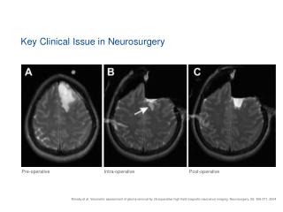
Key Clinical Issue in Neurosurgery Pre-operative Intra-operative Post-operative Nimsky et al. Volumetric assessment of glioma removal by intraoperative high-field magnetic resonance imaging. Neurosurgery, 55: 358-371, 2004
iMRI Clinical Applications A B C D Epilepsy fMRI DTI Spectroscopy E F G H Tumor Resection Vascular Surgery Catheter Placement Biopsy Images A - G are courtesy of R. Fahlbusch and Ch. Nimsky, Erlangen-Nuremberg University, Germany Image H is courtesy of L. Ferrante and L. Mastronardi, Ospedale Sant Andrea Rome, Italy
Integration of Navigation Improves Clinical Outcomes “Our results demonstrate that the optimal usefulness of intraoperative technology comes with the combination of iMRI with updated neuronavigation, resulting in both increased average percentage of tumor resection (98%) and decreased residual 3.3 cm³ for resection of supratentorial gliomas. “ Source: Marvin Bergsneider, M.D. et al. Clinical Neurosurgery Chapter 52: 219-222, 2004 Study based on 45 patients with supratentorial gliomas