Introduction to SAXS at SSRL
310 likes | 580 Vues
Everything You Ever Wanted to Know About But Were Afraid to Ask. SAXS. Introduction to SAXS at SSRL. John A Pople Stanford Synchrotron Radiation Laboratory, Stanford Linear Accelerator Center, Stanford CA 94309. When should I use the Scattering Technique?. Ideal Studies for Scattering.

Introduction to SAXS at SSRL
E N D
Presentation Transcript
Everything You Ever Wanted to Know About But Were Afraid to Ask SAXS Introduction to SAXS at SSRL John A Pople Stanford Synchrotron Radiation Laboratory, Stanford Linear Accelerator Center, Stanford CA 94309
When should I use the Scattering Technique?
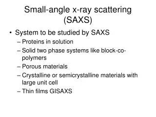
Ideal Studies for Scattering Scattering good for: • Global parameters, distributions; 1st order • Different sample states • In-situ transitional studies • Non destructive sample preparation Solid Melted & Sheared Recrystallized
Ideal Studies for Microscopy Microscopy good for: • Local detail • Surface detail • Faithfully represents local complexities E.g. if objective is to monitor the degree to which Mickey’s nose(s) and ears hold to a circular micromorphology… use microscopy not scattering
Complementary Scattering and Microscopy 200 nm 5 mins in conc HNO3 60 mins Forming a bi-continuous porous network with ligament width on the nanoscale by removing the less noble element from a binary alloy, in this case Ag-Au
Scattering: Neutrons or Photons? X-rays Sensitive to electron density contrast Neutrons Sensitive to nuclear scattering length contrast Neutron scattering: Deuteration allows species selection X-ray scattering: Relatively small sample quantities required Relatively fast data acquisition times - allows time resolved effects to be characterized
Scattering: Neutrons or Photons? Neutrons: Deuteration allows species selection This essentially permits a dramatic alteration to the ‘visibility’ of the tagged elements in terms of their contribution to the reciprocal space scattering pattern Atom Scattering length Incoherent scattering (x 1012 cm2) (x 1024 cm2) 1H -0.374 80 2D 0.667 2
Scattering: Neutrons or Photons? Photos of deformation SANS patterns l= 0% l= 300%
Scattering: Neutrons or Photons? X-rays: Order of magnitude better spatial resolution Fast data acquisition times for time resolved data Oscillatory Shearing of lyotropic HPC – a liquid crystal polymer
X-ray Scattering: Transmission or Reflection? Need to be conscious of: Constituent elements, i.e. absorption cutoffs Multiple scattering Area of interest: surface effect or bulk effect • Transmission geometry appropriate for: • Extracting bulk parameters, especially in deformation • Weakly scattering samples: can vary path length
X-ray Scattering: Transmission or Reflection? • Reflection geometry appropriate for: • Films on a substrate (whether opaque or not) • Probing surface interactions
No fundamental difference in physics: a consequence of chemistry X-ray Scattering: SAXS or WAXS? WAXS patterns contain data concerning correlations on an intra-molecular, inter-atomic level (0.1-1 nm) SAXS patterns contain data concerning correlations on an inter-molecular level: necessarily samples where there is macromolecular or aggregate order (1-100 nm) As synthesis design/control improves, SAXS becomes more relevant than ever before
Experimental consequences X-ray Scattering: SAXS or WAXS? • WAXS: Detector close to sample, consider: • Distortion of reciprocal space mapping • Thermal effects when heating sample • No ion chamber for absorption • SAXS: Detector far from sample, consider: • Absorption from intermediate space • Interception of appropriate q range
Recognizing Reciprocal Space Patterns: Indexing Face centered cubic pattern from diblock copolymer gel
≡1; =√2; =√3 ≡1; =√3; =√4 ≡1; =√4/3; =√8/3 Recognizing Reciprocal Space Patterns: Indexing Real space packing Hexagonal Body centered cubic Face centered cubic Reciprocal space image (unoriented domains) Normalized peak positions
Real space packing Recognizing Reciprocal Space Patterns: Preferential Orientation Reciprocal space image Hydrated DNA Preferentially aligned rods Randomly aligned rods
Extracting Physical Parameters from X-ray data q f I(q) I(f) q f
ln I(q) q2 Molecular size: Radius of gyration (Rg) Extracting Physical Parameters from X-ray data I(q) = I(0) exp [-q2Rg2 / 3] Rg2a ln I(q) / q2 Guinier plot Guinier region: q < 1 / Rg
q-1 Rod Coil in good solvent q-5/3 q-4 Sphere Molecular conformation: Scaling exponent Extracting Physical Parameters from X-ray data Gradient of profile in intermediate region implies fractal dimension of scattering unit Guinier plateau ln I(q) Intermediate region ln q
DEJ pulp John H Kinney Department of Preventive and Restorative Dental Sciences, University of California, San Francisco, CA 94143 Molecular Conformation in Dentin q Q SAXS pattern
plate-like needle-like DEJ pulp Molecular Conformation in Dentin Shape change of mineral crystallites from needle-like to plate-like from pulp to dentin-enamel junction (DEJ). Dentinogenesis imperfecta (DI) teeth shown to exhibit impaired development of intrafibrillar mineral: characteristic scattering peaks are absent from the diseased tooth.
I(q) q2 q* q Molecular conformation: Persistence length of coiled chain Extracting Physical Parameters from X-ray data Kratky plot persistence length = 6 / (p q*)
I(f) q f f Molecular orientation: Orientation parameter P2 Extracting Physical Parameters from X-ray data <P2n(cos f)> = I(s,f) P2n(cos f) sin f df I(s,f) sin f df Normalized: -0.5 < P2 < 1 Azimuthal profile
Molecular Orientation in Injection Moldings Measuring the degree and inclination of preferential molecular orientation in a piece of injection molded plastic (e.g. hip replacement joints). ~ 1500 WAXS patterns Marks the injection point Axis of orientation Orientation parameters: 0 < P2 < 0.3
SSRL Beamline 1-4: SAXS Materials Science ion chambers shutter X-rays N2 supply beam defining slits sample stage guard slits CCD detector optical rail & table
Rheology of Straight and Branched Fatty Alcohols Linear Eicosanol increasing surface pressure: 0 to 40 mN/m q (/nm) Study phase transitions of Langmuir monolayers of mixed fatty alcohols in terms of molecular branching and surface tension linear eicosanol (C20H42O, MW = 298) branched eicosanol (C20H42O, MW = 298) Surface tension sensor Gold mirror x-ray path Langmuir trough