The Endocrine System
The Endocrine System. Chapter 25. The Endocrine System. A system of ductless glands that secrete hormones (‘messenger’) molecules - secrete hormones directly into the blood or lymph - hormones trigger physiological changes in target cells


The Endocrine System
E N D
Presentation Transcript
The Endocrine System Chapter 25
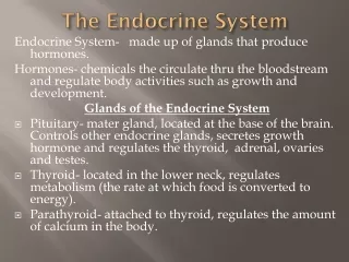
The Endocrine System • A system of ductless glands that secrete hormones (‘messenger’) molecules - secrete hormones directly into the blood or lymph - hormones trigger physiological changes in target cells • Controls and integrates the functions of other body systems - closely interacts with the nervous system • Endrocrinology - the study of hormones and endocrine glands
Nervous System Controls homeostsis rapidly Anatomically continuous: nerve impluse conducted along axons from one neuron to the next Neurotransmitters (NTs) Brief effect (muscle contraction) Endocrine System Controls growth and metabolism slowly Scattered: messenger molecules released into the EC space will immediately enter adjacent capillaries Hormones (‘to excite’) Longer lasting effect with feedback loops Together these systems interact to coordinate and integrate activity of our cells
Endocrine Organs • Series of ductless glands - small and scattered throughout the body - some may be both endocrine and exocrine • ‘Pure’ endocrine glands - pituitary, pineal, thyroid, parathyroid, and adrenal • Organs containing endocrine cells - pancreas, thymus, gonads, and hypothalamus • Most endocrine cells are of epithelial origin - others include hormone-secreting neurons, muscle cells, and fibroblast-like cells • Highly vascularized – blood and lymph vessels
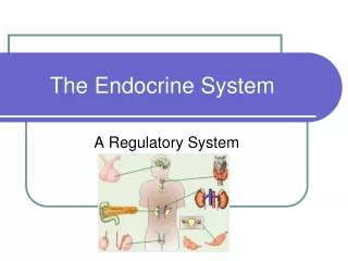
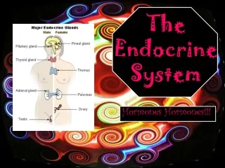
Location of the Major Endocrine Glands Endocrine cells also occur in the heart, alimentary canal, kidney, skin, placenta, and elsewhere Fig 25.1
Endocrine System Overview Endocrine glands may be stimulated by the nervous system or chemical changes in the body - respond by secreting hormones into the circulation Hormones travel through the bloodstream but affect only specific tissues called target tissues Hormones secreted by cells regulate the metabolic function of other cells in the body - effects result from pre-programmed responses of target cells
Endocrine Functions • Regulation of: - Internal environment (adjust fluid/volume ratio):aldosterone - Metabolism and energy balance: thyroid hormones - Cardiac and smooth muscle contraction: epinephrine and norepinephrine - Immune system: cytokines - Glandular secretions: hypothalamus and pituitary hormones • Maintainance and assistance of: - Homeostasis despite disruptions: pancreatic hormones - Growth and development: growth hormones - Reproduction: hormones that influence oogenesis and spermatogenesis
Classes of Hormones The body produces many different kinds of hormones with distinct chemical structures Two broad molecular categories: 1. Amino acid-based hormones - modified amino acids (amines), peptides (short chains of amino acids), and proteins (long chains of amino acids) 2. Steroid hormones - lipid molecules derived from cholesterol
Basic Hormone Action • Hormones circulate throughout the body in BVs - leave the bloodstream at capillaries encountering all body tissues - influences only specific tissue cells or target cells - same hormone can have different effects on different target cells - similar molecular structures can have very different functions • Cells have receptors on their surface that bind only specific types of hormones - receptor binding initiates a response/reaction - hormones are just molecular triggers and do not carry any coded information
Control of Hormone Secretion Secretion is triggered by three major types of stimuli: humoral, neural, and hormonal stimuli Humoral (‘body fluids’) - simplest of endocrine control mechanisms - secretion is in direct response to changing critical ion or nutrient levels in the blood - parathyroid monitors calcium: responds to decline be secreting hormone to reverse decline Neural - a few glands secrete their hormones in response to stimuli by the nervous system to induce physiological changes - sympathetic nerve fibers stimulate cells in the adrenal medulla - induces release of epinephrine and norepinephrine
Control of Hormone Secretion • Hormonal - many endocrine glands secrete their hormones in response to hormonal stimuli received from other endocrine glands - certain hormones signal secretion of other hormones - the hypothalamus secretes hormones stimulates the pituitary stimulates other glands (the thyroid, adrenal cortex, and the gonads) Note: the hypothalamus is called the master gland - controls many functions of the endocrine system, through hormonal and other mechanisms
Control of Hormone Secretion • Always controlled by feedback loops - ensures that hormone concentrations stay within a narrow ‘desirable’ range in the blood • Negative feedback loop: - Hormonal blood concentration declines below a minimum set point more hormone is secreted - Blood concentration exceeds a maximum set point hormone production is halted • Positive feedback loop: - as blood concentrations of a certain hormone increase the response of the effector organ stimulates further secretion - progression of labor in childbirth by oxytocin
The Pituitary Gland (Hypophysis) The pituitary gland (hypophysis - undergrowth): - secretes 9 major hormone - sits in the hypophyseal fossa of the sella turcica - resembles a golf club: the gland forms the head of the club, and the stalk, called the infundibulum (funnel), forms the shaft - the infundibulum connects superiorly to the hypothalamus 2 basic divisions of the pituitary gland: - Anterior adenohypophysis (adeno = glandular) - Posterior neurohypophysis (neuro = neural) Blood supply: 2 branches of the internal carotid artery - superior hypophyseal artery supplies the adenohypophysis - inferior hypophyseal artery supplies the pars nervosa
The Pituitary Gland Figure 25.3a–c
The Adenohypophysis Hormone release is controlled by the hypothalamus - stimulated and inhibited by releasing and inhibiting hormones 3 subdivisions: pars distalis, pars intermedia, pars tuberalis - pars distalis the largest division has 5 different endocrine cell types - secrete protein hormones, have many secretory granules and a well-developed RER and Golgi apparatus Somatotropic cells - secrete growth hormone (GH) Mammotropic cells - secrete prolactin (PRL)
Pars Distalis Endocrine Cells • Thyrotropic cells - secrete thyroid-stimulating hormone (TSH) • Corticotropic cells - secrete adrenocorticotropic hormone (ACTH) and melanocyte-stimulating hormone (MSH) • Gonadotropic cells - secrete follicle-stimulating hormone (FSH), luteinizing hormone (LH) • Tropic (‘changing’) hormones – TSH, ACTH, FSH, LH - regulate secretion of hormones by other endocrine glands - GH, PRL, and MSH act directly on nonendocrine target tissues • 5 cell types group into 3 categories when stained: acidophils, basophils, chromophobes
GH or somatotropin- stimulates growth of body tissues, especially in muscle and skeleton • TSH -stimulates the thyroid to produce and release T4 and T3 (influences metabolism) • PRL– initiates and maintains milk production (lactation) by mammary glands in the breasts • ACTH– influences production and secretion of hormones by the adrenal cortex (helps us deal with stress) • MSH- stimulates melanocytes of the epidermis to produce more melanin, thus darkening the skin • FSH- stimulates ova maturation and estrogen production in ovaries and sperm production in the testes • LH or ICSH– stimulates ovulation and progesterone secretion in ovaries and testosterone secretion in the testes
Hypothalamic Control of Hormone Secretion from the Adenohypophysis • The hypothalamus have neurons that produce and release hormones much like NTs are released - secretes releasing factors to release hormones - secretes inhibiting hormones to turn off hormone secretion - travels through the hypophyseal portal system into the anterior pituitary to stimulate its hormone secretion - the hypophysial portal system involves two beds of capillaries connected by a vein - allows a high level of hormone concentration within a small region - designed so that the hormones released by the hypothalamus travel directly to the anterior pituitary - in turn the anterior pituitary releases hormones into systemic circulation
Hypothalamic Control of Hormone Secretion from the Adenohypophysis Fig 25.4
The Neurohypophysis • Stores and releases hormones produced by the hypothalamus - structurally part of the brain - composed of nervous tissue (unmyelinated axons and neuroglial cells) • Hormones made in the neuron cell bodies, transported along the axons, and stored in dilated axon terminals (Herring bodies) – secrete 2 hormones - antidiuretic hormone (ADH) or vasopressin (‘vessel constrictor’): targets the kidney to resorb more water from the urine and return it to the blood - secretes oxytocin induces contractions of smooth muscle of reproductive organs
The Neurohypophysis When the neurons fire, they release stored hormones into a capillary bed in the pars nervosa for distribution Fig 25.5
The Thyroid Gland • Largest pure endocrine gland - located in the anterior neck • Internally, is composed of hollow follicles - separated by areolar CT rich in capillaries - walls are formed of cuboidal or squamous epithelial cells (follicular cells) - lying within the epithelium are parafollicular (C) cells - central lumen filled with colloid (‘gluelike’) consisting of thyroglobulin (protein precursor to thyroid hormone) • Produces 2 hormones: amino-based and protein - Thyroid hormone (TH): thyroxine (T3 ); tri-iodothyronine (T4) - Calcitonin: lowers blood levels of Ca2+, mostly during childhood
The Thyroid Gland T4 and T3, consists of 2 amino acids and iodine Main function is to increase metabolic rate Fig 25.6
Histology of the Thyroid Gland • Follicle cells continuously synthesize thyroglobulin and secrete it into the follicle lumen for iodination and storage • TSH (pituitary gland) signals the follicle cells to release TH Fig 25.6
The Parathyroid Glands • Lie on the posterior surface of the thyroid gland surrounded by CT capsules (number varies) • Contains thick branching cords composed of 2 types of endocrine cells - small abundant chief cells and rare larger oxyphil cells • Chief cells produce a small protein hormone, PTH - PTH increases calcium levels and is essential to life: 1) stimulates osteoclasts to release calcium from bones 2) decreases secretion of calcium by the kidney 3) activates vit D, which stimulates uptake of Ca by the intestine
The Parathyroid Glands Posterior view of the pharynx a and trachea showing the location of the parathyroid glands on the posterior aspect of the thyroid Fig 25.7
The function of oxyphil (‘acid-loving’) cells is unknown PTH is essential to life - low Ca2+ levels lead to lethal neuromuscular disorders What is the antagonist of PTH? Histology of the Parathyroid Gland Fig 25.7
The Adrenal (Suprarenal) Glands • Paired pyramidal organs on the superior surface of the kidneys – highly vascularized • 3 groups of 60 small suprarenal arteries supply each gland - the superior suprarenal arteries from the inferior phrenic artery; - middle suprarenal arteries from the aorta; - inferior suprarenal arteries from the renal artery • Veins - left suprarenal vein drains into the renal vein and the right suprarenal vein drains into the inferior vena cava
The Adrenal (Suprarenal) Glands • Nerve supply is almost entirely sympathetic fibers • 2 endocrine glands in one - internal and external: - Adrenal medulla: a cluster of neurons derived from the neural crest, acts as part of the sympathetic NS - Adrenal cortex: bulk of the adrenal gland derived from mesoderm
The Adrenal Medulla • Part of the autonomic nervous system (Ch 15) • Chromaffin (‘affinity for chromium’) cells - arranged in spherical clusters with some branching cords - modified ganglionic sympathetic neurons - secrete catecholamines: the amine hormones epinephrine and norepinephrine - active in the ‘fight, flight, and fright’ (fight or flight) response - hormones stored in secretory vesicles
The Adrenal Cortex • Secretes a variety of corticosteroid hormones - all are lipid-based steroids • Cortex is composed of 3 layers or zones: - Zona glomerulosa (‘ball of yarn’): cell clusters - Zona fasciculata: cells arranged in bundles - Zona reticularis (‘network’): cells arranged in a branching network
The Adrenal Gland Figure 25.8a, b
Adrenal Corticosteroids • 2 main classes: mineralocorticoid & glucocorticoid • Main mineralocorticoid is aldosterone - secreted by the zona glomerulosa - in response to a decline in blood volume or BP - prompts kidney to resorb more sodium into the blood; water passively follows, increasing blood volume
Adrenal Corticosteroids • Glucocorticoids: cortisol is the main type - secreted by zona fasciculata and zona reticularis - helps the body deal with stressful situations by keeping glucose levels high to support the brain - body cells switch to fats and amino acids as energy sources - high amounts depress the inflammatory response
Adrenal Corticosteroids • Hormonal pathway of stress: - hypothalamus sends corticotropin-releasing hormone (CRH) to the adenohypophysis, which secretes ACTH - ATCH travels to the adrenal cortex to signal glucocorticoid secretions - the sympathetic NS can also stimulate glucocorticoid secretions
Adrenal Corticosteroids • Zona reticularis secretes an androgen hormone, dehydroepiandrosterone (DHEA) - DHEA is converted to testosterone and estrogens in peripheral tissues - proposed beneficial effects include counteracting stress, boosting immunity, and mood
Structure of Steroid-Secreting Cells • Steroid-secreting cells have distinctive features - abundant SER and no secretory granules - mitochondria have unusual cristae shaped like tubes - lipid droplets are abundant in cytoplasm (lipids = raw material of steroids) • Characterize cells of the adrenal cortex - also testicular and ovarian cells which secrete steroid sex hormones: the interstitial cells, theca folliculi cells, and cells of the corpus luteum
Interstitial cell in the testis Fig 25.9
The Pineal Gland • Small, pine cone shaped structure at the end of a short stalk on the roof of the diencephalon • Pinealocytes are arranged in both spherical clusters and branching cords - star-shaped cells with long, branching cell process - dense particles of calcium lie between the cell clusters, forming the ‘pineal sand’ (which is radiopaque) - in Xrays used as a landmark to identify brain structures - secretes melatonin: a hormone that regulates circadian rhythms (hypothalamus responds to a lack of visual input)
The Pancreas • Located in the posterior abdominal wall • Contains endocrine and exocrine cells • Exocrine acinar cells, form most of the gland - secrete digestive enzymes into the small intestine • Endocrine cells are contained in spherical bodies - pancreatic islets or islets of Langerhans - about 1 million scattered among the exocrine acini
The Pancreas • Main endocrine cell types: - Alpha cells (α cells): secrete glucagon signals liver to release glucose from glycogen; raises blood sugar - Beta cells (β cells): secrete insulin signals most body cells to take up glucose from the blood; promotes glucose storage as glycogen in liver; lowers blood sugar
A Pancreatic Islet Figure 25.10
The Thymus • Located in the lower neck and anterior thorax • Important immune organ • Site at which T-lymphocytes arise from lymphocyte-precursor cells - transformation stimulated by thymic hormones, secreted by the thymus epithelial reticular cells • Thymic hormones – a family of peptide molecules, including thymopoietin and thymosin
The Gonads • Main sources of sex hormone – testes and ovaries • Male testes - interstitial cells secrete androgens (primarily testosterone) - promotes the formation of sperm - maintains secondary sex characteristics
The Gonads • Female ovaries - androgens secreted by the theca folliculi directly converted into estrogens by the follicular granulosa cells & progesterone - estrogens and progesterone secreted by the corpus luteum - estrogens maintain reproductive organs and secondary sex characteristics - progesterone signals uterus to prepare for pregnancy
Endocrine cells occur within The heart - the atria contains atrial natriuretic peptide (ANP) - hormone that stimulates the kidneys to produce more urine containing salt - getting rid of the excess fluid and salt reduces excess blood volume and salt levels; reduces blood pressure The GI tract has scattered enteroendocrine cells - release amino acid/peptide hormones chemically similar to neurotransmitters - affect functions related to regulating digestion, blood chemistry, and blood flow Other Endocrine Structures