Download
1 / 52
520 likes | 591 Vues
This resource provides information on audiology tests, hearing loss causes, example audiograms, and more for professionals in Deaf Education, Audiology & Speech Pathology. Quick look-up guide with reference links.

E N D
Audiology Resource Notebook • This presentation is intended as a resource for professionals in Deaf Education, Audiology, and Speech Pathology. It includes information about the basic audiometric test battery, common causes of hearing loss, and example audiograms. Each topic has its own table of contents, and, where possible, each section of information has an accompanying graphic. The information contained within the presentation is meant to be a quick look-up guide to the most essential information with regards to these topics. The reference links at the end of the presentation provide more in-depth information.
Audiology Resource Notebook Katrina García The University of Tulsa Dr. Sharon Baker 2002
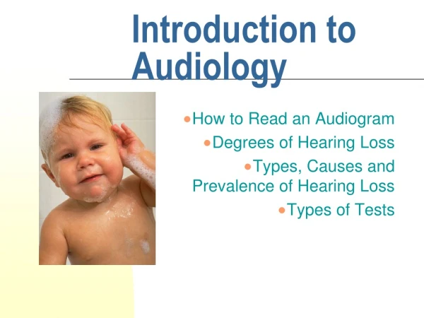
Resource Sections • Audiometric Procedures • Otoscopic Exam – Speech Audiometry • Pure Tone Testing – Immittance Testing • Example Audiograms • Student Preformed Audiogram • Types of Hearing Loss • Audiograms That Indicate a Disorder • Causes of Hearing Loss • Table of Contents
Otoscopic Exam • Using an otoscope, examine the eardrum for any abnormalities. Normal eardrum TM Perforation Serous Otitis Media Return to Resource Sections
Pure Tone Testing Air Conduction • Test right ear or better ear first. • Begin at 1000Hz at 40dB. • Decrease 10dB until there is no response. • Go up 5dB to zero in. • Threshold is established when client responds 50% of the time. • O indicates right ear • Xindicates left ear
Bone Conduction Procedure is the same. < indicates right ear. >indicates left ear. Air conduction indicates amount of loss. Bone conduction indicates type of loss. Air Bone Gap If there is no air bone gap, and there is loss, then the loss is sensorineural. If there is an air bone gap of more than 10dB, and bone conduction is normal, it is a conductive loss. If there is an air bone gap of more than 10dB and bone conduction and air conduction both indicate a loss, the loss is mixed. Pure Tone Testing Continued Return to Resource Sections
Speech Audiometry Speech Reception Threshold (SRT) • Spondees are the speech stimuli. • Start at 40dB, ask client to repeat spondee. • Establish threshold using the down 10dB, up 5dB method. • Compare to the average of the pure tone values at 500, 1000, and 2000Hz. If there is a sharp decline in one value, use the best two. • Should be within 15dB of Pure Tone Average (PTA). If not, suspect a problem with the pure tone results. • A spondee board can be used for inarticulate clients. • Speech Awareness Threshold (SAT) is used when client is deaf. In this case, a repeated syllable is used and the client is asked to indicate when they can perceive the sound.
Speech Audiometry Continued Word Recognition Score (WRS) • The stimuli are a list of 25-50 phonetically balanced one syllable word. • Present every word at the same loudness and say it only once. • Ask client to repeat word after it is said. • Use 25-50 words for each ear and mark how many words are repeated correctly. • Calculate percentage of words recognized. 100-96% is excellent. Return to Resource Sections
Immittance Testing Tympanometry • The tympanogram is a plot of middle air pressure vs. TM compliance. • There are three types of tympanogram: Type A Type B Type C
Immittance Testing Continued • Type A is normal, and indicates the mobility of the ear drum. • Type A has two subtypes. Type AD indicates a great deal of flexibility. Type As can indicate that the eardrum’s movement is being restricted. • Type B indicates fluid in the middle ear. • Type C indicates low air pressure in the middle ear. • The eustachian tubes are not letting enough air through • Can lead to a fluid filling the ear. Return to Resource Sections
Student Performed Audiogram • Performed 12-6-01 • Client: E. S., age 17 (DOB 12-28-83) • Examiner: Katrina García • SAT: 80dB, 75dB Comments: Client has a bilateral mixed loss with recruitment that is moderately severe to profound. SAT is consistent with audiogram. Cause of deafness is unknown and most likely congenital. Return to Resource Sections
Conductive Loss Return to Pure Tone Testing
Sensorineural Loss Return to Pure Tone Testing
Mixed Loss Return to Pure Tone Testing Return to Resource Sections
Audiograms That Indicatea Disorder • Otosclerosis • Presbycusis • U-Shaped • Treacher Collins • Rubella • Noise-Induced Hearing Loss Return to Resource Sections
Noise-Induced Hearing Loss Noise-Induced hearing loss is indicated by high frequency loss usually of 4000Hz or higher Return to Audiograms
Disorders/Conditions Atresia Ceruminosis TM perforation Barotrauma Otosclerosis Presbycusis Cleft Palate Multiple Sclerosis Infections Otitis externa Otitis media Labyrinthitis Tumors Cholesteatoma Vestibular Schwannoma Table of Contents
Syndromes Alport syndrome Down syndrome Waardenburg syndrome Usher syndrome Treacher Collins syndrome Meniere’s disease TORCH Complex Toxoplasmosis Congenital Syphilis Rubella Cytomegalovirus Herpes Simplex Table of Contents Continued Previous Page Return to Resource Sections
Atresia is the absence of an external ear canal. Is almost always accompanied by abnormalities in the outer and middle ear. In most cases there is a bony plate the separates the external ear from the contents of the middle ear. Conductive loss, bone conduction hearing aids are used. Can be surgically corrected in some instances. Atresia Atresia without microtia. Return to Table of Contents
Buildup of the wax (cerumen) in the outer ear canal. Can be caused by over- productive cerumen glands, but is most often caused when attempts to clean the ear pushes wax deeper into the canal Causes conductive hearing loss. Can usually be cleared up with drops that soften the wax. Complications that can occur include: TM perforation, otitis media, otitis externa, and permanent hearing loss. Cerumenosis Return to Table of Contents
A hole or rupture in the eardrum. Causes conductive hearing loss, the larger the perforation the greater the loss Pain is usually not present. Causes usually trauma or infection. Picture of TM perforation due to trauma If caused by otitis media, there may be infected or bloody drainage. Most perforations will heal spontaneously within weeks of rupture. During healing, ear must be protected from water and trauma On rare occasions a small hole may remain after PE tubes have fallen out or been removed TM Perforation Return to Otoscopic Exam Return to Table of Contents
Discomfort in the ear caused by pressure differences between the outer and middle ear. Barotrauma can occur when flying, scuba diving, and changing altitude. Symptoms include pain, slight conductive hearing loss, and ear fullness If severe or prolonged, can cause moderate to severe hearing loss, feeling of pressure in the ears, nosebleed and middle ear pain. Otoscopic exam may show slight bulge or retraction of the eardrum. Severe barotrauma may be hard to distinguish from otitis media. Barotrauma Return to Table of Contents
Otosclerosis is the growth of spongy bone-like tissue around the footplate of the stapes and blocks the oval window. There are several types: Subclinical does not interfere with the ossicles Histologic may or may not cause hearing loss Clinical can be present in teen years, but may not be noticed until adulthood Causes gradual conductive hearing loss progressing from the low frequencies, to the high frequencies, and then to the middle frequencies. Can also cause sensorineural loss in the high frequencies, this type is known as cochlear otosclerosis. Can begin unilaterally and progress to a bilateral loss Otosclerosis
Maximum conductive loss is between 50-60dB, with a mixed loss it will increase to 60-70dB. Usually occurs between the ages of 30 and 50, but can occur as early as late childhood. Women are more likely to develop a hearing loss from otosclerosis than men. Women may notice a decrease in hearing after pregnancy due to hormonal changes. Most likely has a genetic component. Conductive loss can be corrected with a stapedectomy which replaces the damaged stapes Otosclerosis Continued
Audiogram: Otosclerosis One of the characteristic features of an audiogram that indicates otosclerosis is a smaller air bone gap at 2000Hz. This is known as a Carhart notch. Return to Audiograms Return to Table of Contents
Hearing loss associated with the degenerative effects of aging. It affects 30-35% of adults between 65 and 75 and increases to 40-50% of those over 75 Loss is sensorineural and in the high frequencies Tinnitus may occur There is a central auditory component that will result in lower WRS than expected Client reports that the speech of others sounds slurred or mumbled Conversations are harder to hear when there is background noise. Presbycusis
Audiogram: Presbycusis Audiograms that indicate presbycusis show a sharp drop off in the frequencies above 2000Hz. Return to Audiograms Return to Table of Contents
Cleft Palate • Children with a cleft palate have more ear and sinus infections • Causes conductive hearing loss due to improper drainage of the middle ear • Chronic otitis media can result in a language delay • 5% of cases occur with syndromes, including Treacher Collins Bilateral cleft palate and cleft lip Return to Table of Contents
Multiple Sclerosis (MS) • 6% of MS patients experience hearing loss • In rare cases hearing loss is the first symptom of MS • It causes a fluctuating sensorineural loss • Hearing loss may take place when MS flares • Results from an ABR will not be consistent or repeatable Return to Table of Contents
Otitis Externa • Inflammation of the ear canal, also known as swimmer’s ear. • Can be caused by fungi, mold, or bacteria • Symptoms include pain, itching, tender tragus, fever, swelling, and discharge
Occurs in elderly diabetic patients, can occur in AIDS patients Characterized by persistent and severe earache, foul-smelling purulent otorrhea, and granulation tissue in the ear canal. Caused by the bacteria Pseudomonas aeruginosa Causes varying degrees of conductive hearing loss Effects the facial nerve in severe cases Has a mortality rate of about 50% Malignant Otitis Externa Return to Table of Contents
Otitis Media • Inflammation of the middle ear • Most common cause is eustachain tube dysfunction • Occurs most often in children under 3 years of age because of horizontal position of eustachain tubes • Occurs as a complication of a cold, sore throat, or other upper respiratory infections • Can occur with or without fluid (effusion) • Effusion can occur with or without infection
Otitis Media Continued • Serous otitis media is characterized by thing watery fluid that is free from infection • Symptoms include ear fullness and hearing loss • Suppurative otitis media is characterized by fluid that contains puss. It is indicative of infection. • Symptoms include fever, ear pain, hearing loss, ear fullness, dizziness, nausea, and bulging red or yellow eardrum Return to Otoscopic Exam
Otitis Media Continued Mucoid fluid is sometimes seen with a low grade infection. Adhesive otitis media, or glue ear, is characterized by a thick, sticky effusion that causes the eardrum to become severely retracted. Return to Table of Contents
Inflammation of the membranous labyrinth and semicircular canals of the inner ear Can occur following otitis media, or an upper respiratory infection Symptoms include tinnitus, dizziness, vertigo, nausea, loss of balance, and hearing loss Hearing loss and vestibular impairments caused by bacterial labyrinthitis are often permanent The damage caused by viral labyrinthitis may or may not be permanent Labyrinthitis Return to Table of Contents
Cholesteatoma • A collection of skin, skin tissue, and debris in the epitympanic recess of the middle ear • Is associated with chronic otitis media and TM perforation • Symptoms include foul smelling otorrhea, middle ear pressure, and hearing loss.
Cholesteatoma Continued • Cholesteatoma can be congenital, but is most often associated with ear infections • Causes conductive hearing loss • Can erode ossicular chain • Can be life-threatening because it can erode bone between the middle ear and the brain, causing meningitis • Must be removed surgically Return to Table of Contents
A tumor on the Schwanncells of the vestibular portion of the auditory nerve Usually starts in the internal auditory canal Very slow growing Unilateral 95% of the time Symptoms include hearing loss, tinnitus, vertigo, and facial numbness and twitching Hearing loss is due to compression or infiltration of auditory nerve Loss begins with the high frequencies Can lead to total deafness in the tumor ear Atypical forms of hearing loss are common SRT is out of proportion to pure tone results Vestibular Schwannoma
Vestibular Schwannoma Continued Small Vestibular Schwannoma Itracanalicular Vestibular Schwannoma Large Vestibular Schwannoma Return to Table of Contents
Genetic condition that is X-linked (85%), Autosomal recessive (15%), and autosomal dominant (<1%) Symptoms include renal disease, eye disorders, blood platelet defects, and bilateral sensorineural hearing loss Hearing loss can be progressive and delayed in onset Loss is usually mild to moderate Usually diagnosed by testing for blood or protein in urine Alport Syndrome Return to Table of Contents
Down Syndrome • Caused when a child has three #21 chromosomes instead of two • Symptoms include characteristic facial features, congenital heart disease, upper respiratory infections, mental retardation of varying degrees, recurrent otitis media, and conductive or mixed hearing loss • 60-80% of children with Down Syndrome experience hearing loss • Life expectancy of a person with Down Syndrome is 55 years Return to Table of Contents
There are four types of Waardenburg Syndrome It is autosomal dominant Symptoms include wide set eyes, white forelock that can extend to the skin, different pigmentation in each eye, and sensorineural hearing loss Type I has a 20% chance of non-progressive sensorineural hearing loss both unilaterally and bilaterally that is mild to profound Type II has a 50% higher chance for unilateral sensorineural loss Waardenburg Syndrome
Audiogram: Waardenburg Characteristic audiogram shapes include low frequency U shaped losses, and sometimes a combination of low frequency sensorineural loss in one ear and profound loss in another Mild to moderate U-shaped loss Return to Table of Contents Return to Audiograms
Usher Syndrome • Autosomal recessive • Characterized by moderate to profound sensorineural loss with progressive vision loss from Retinitis pigmentosa • Hearing loss can be congenital (severe to profound) or progressive • Types of blindness include night blindness, difficulty adjusting to changes in light conditions, and tunnel vision • Children with these types of losses may at first be described as clumsy Return to Table of Contents
Treacher Collins • Autosomal dominant • Characterized by facial nerve abnormalities, craneofacial anomalies including outer and middle ear deformities (microtia, atresia, preauricular tags or pits, stenosis) • Mental function is normal • Hearing loss is conductive or mixed Return to Table of Contents
Meniere’s Disease • Characterized by: • episodic vertigo • roaring tinnitus • sensation of ear fullness • fluctuating and progressive low-frequency sensorineural hearing loss • May be caused by endolymphatic hydrops • Is rarely reported in children Return to Table of Contents
Parasitic infection transmitted across the placenta Can be present in raw meet, raw eggs, and cat feces Greatest risk to baby during the first trimester Mother is asymptomatic Causes congenital sensorineural loss that is mild-profound As much as 10-20% of congenital deafness is contributed to toxoplasmosis Also causes mental retardation, hydrocephalus, vision loss, seizures, and neuro-muscular problems Can be treated with antiparasitic drugs both prenatally and postnatally Toxoplasmosis Return to Table of Contents
Bacterial sexually transmitted disease Mother can be treated with antibiotics during pregnancy Child is asymptomatic at birth and may not manifest disease for several decades Hearing loss is extremely varied Audiograms show odd configurations of varying degrees Clients tend to have a much poorer WRS than indicated by audiogram Congenital Syphilis Return to Table of Contents
Rubella • Viral infection the crosses the placenta • Worst if contracted during the first trimester • Mother is asymptomatic • Causes visual cataracts,heart disease, bilateral sensorineural hearing loss, mental retardation Causes a u-shaped loss Return to Table of Contents Return to Audiograms
The most common viral cause of congenital hearing loss Easily transmitted, as much as 80% of the population in the US may be carriers 40-50% of infected mothers will pass CMV on to their infants 90% of infants are asymptomatic at birth, those who are not have a 30% mortality rate Causes vision loss, progressive sensorineural hearing loss, and mental retardation Congenital Cytomegalovirus (CMV) Return to Table of Contents