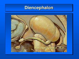
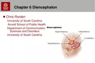
Chapter 6 Diencephalon
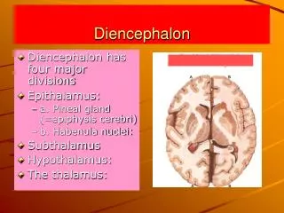
Chapter 6 Diencephalon. Chris Rorden University of South Carolina Arnold School of Public Health Department of Communication Sciences and Disorders University of South Carolina. Diencephalon – Gross Anatomy. Four Parts Thalamus Epithalamus Subthalamus Hypothalamus.

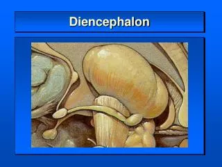
Chapter 6 Diencephalon
E N D
Presentation Transcript
Chapter 6 Diencephalon • Chris Rorden University of South Carolina Arnold School of Public Health Department of Communication Sciences and Disorders University of South Carolina
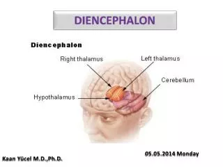
Diencephalon – Gross Anatomy • Four Parts • Thalamus • Epithalamus • Subthalamus • Hypothalamus
Midsagittal Brain Section • Corpus Callosum • Thalamus • Pons
Thalamus: Integrator and gateway for information Subthalamus: Important in motor control Hypothalamus: Mediates endocrine and metabolic states Epithalamus: Diurnal & automatic body functions (diurnal functions refer to regulation of sleep and wakefulness, body temperature, and metabolic rate) Functions
Thalamus • Channels sensory information • pain, taste, temperature, audition, vision • Integrates sensorimotor information • From Basal Ganglia, Cerebellum, and Cortex • Regulates function of association cortex and cortically mediated speech, language, and cognitive functions.
Three levels of Nuclei Medial Lateral Ventral Many Nuclei or receiving groups of neurons on thalamus at each level Thalamic Structure
Related to Limbic Brain and contributes to direction of digestive respiratory urogenital endocrine functions Thalamus: Anterior Nucleus A Anterior nucleus P
Reticular Nucleus Only thalamic nuclei that does not have cortical outputs • Located between external medullary lamina and internal capsule • Receives and projects within thalamus • Integrates and regulates thalamic activity A P Reticular nucleus
Intralaminar Nuclei • Complex in core of internal medullary lamina • Afferent Connection • Globus Pallidus, Vestibular N., Superior colliculus, brainstem reticular formation, Cortex, Brainstem, Cerebellum • Efferent Connection • Basal Ganglia and Cortical Areas • Modulates Excitability of association cortex
Medial Nuclear Complex Dorsomedial nuclei • Dorsomedial Nuclei: • Afferent connections from prefrontal cortex, hippocampus, centromedianum nucleus, hypothalamus • Efferent projections to prefrontal and orbitofrontal cortex and limbic structures • Integrates emotion, thought, and judgment • Destruction lowers threshold for rage • May play a role in Korsakoff's syndrome A P Intralaminar nuclei
Medial Nuclear Complex • Midline N. Complex • Afferent Connections from brainstem reticular formation • Efferent Connections to Cingulate gyrus and hypothalamus • Important in visceral functions
Lateral Nuclear Complex • Dorsolateral N. • Contributes to visceral sensory integration • Lateral Posterior N. • Multisensory Receiving Area • Pulvinar • Connects visual areas with association cortex • Important in language formation, language processing, lexical properties, reading writing • Injury can lead to spatial neglect
Thalamic Anatomy Pulvinar Midline nuclear Complex Lateral posterior nucleus Dorsolateral nucleus
Ventral Nuclear Complex • Ventral Anterior N. • Premotor cortex and skilled movements • Voluntary movements • Ventrolateral N. • Contributes to voluntary motor tasks • Ventral Posterior N. • Sensation from Body and Face
Thalamic Anatomy Ventral Anterior N Ventrolateral N Ventral posterior N
Geniculate Bodies • Lateral Geniculate N • Relay center for Vision • Medial Geniculate N • Relay center for Audition
Epithalamus • Pineal Gland • Cone shaped endocrine (release hormones) structure • Inhibitory influence over gonadal function (sex function) • Diurnal rhythms • Habenular Nucleus • Serves autonomic function and emotional drives
Subthalamus • Subthalamic Nuclei • Motor functions • Hemiballism (motor disorder: involuntary violent movements, persists only during wakefulness) • Zona Incerta • Visuomotor Coordinator
Thalamic Anatomy • Coronal Slice Subthalamus
Cognitive Functions of Thalamus • Involved in language and speech functions • Types of subcortical aphasia include a thalamic based aphasia (left dominant thalamus) • Word Fluency Problems • Neurogenic stuttering from surgeries in thalamus • Thalamic Syndrome • Gross detection of sensations at thalamic level • Thalamic pathologies can result in very strong misinterpretations of sensation
Hypothalamus • Hypothalamus • Near Optic chiasm and Mammillary Bodies • Hypophysis (Pituitary gland) • Hormones regulates body temp, water and food intake, metabolism, sexual behavior, anger, aggression. • E.G. Thyroid Stimulating Hormone