Download

1 / 80
800 likes | 978 Vues
Nerves, Hormones, and Homeostasis. Topic 6.5 Option H1. Nervous System: Structure and Function. Nervous systems are the most intricately organized data-processing systems on Earth. Neuron Nerve cells; your brain contains an estimated 100 billion

E N D
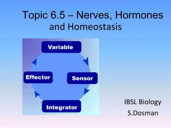
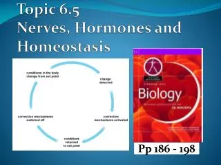
Nerves, Hormones, and Homeostasis Topic 6.5 Option H1
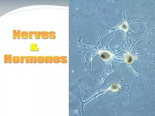
Nervous System: Structure and Function • Nervous systems are the most intricately organized data-processing systems on Earth. • Neuron • Nerve cells; your brain contains an estimated 100 billion • Specialized for carrying signals from one location in the body to another. • consists of a cell body, containing the nucleus and cell organelles, and long, think extensions called neuron fibers that convey signals. • Each neuron may communicate with thousands of others, forming networks that enable us to learn, remember, percieve our surroundings, and move.
Nervous System: Structure and Function • Nervous system has two main divisions: • Central nervous system (CNS) • Consists of the brain and, in vertebrates, the spinal cord • Peripheral nervous system (PNS) • Made up mostly of communication lines called nerves that carry signals into and out of the CNS • A nerve is cable-like bundle of neuron extensions tightly wrapped in connective tissue. • In addition to nerves, the PNS also has ganglia, clusters of neuron cell bodies.
Nervous System: Structure and Function • A nervous system has three interconnected functions: • Sensory input • The conduction of signals from sensory receptors, such as light-detecting cells of the eye, to integration centers. • Integration • The interpretation of the sensory signals and the formulation of responses. • Motor output • The conduction of signals from the integration centers to effector cells, such as muscle cells or gland cells, which perform the body’s responses.
Nervous System: Structure and Function • The relationship between neurons and the nervous system structure and function is easiest to see in the relatively simply circuits that produce reflexes, or automatic responses to stimuli • Three functional types of neurons correspond to a nervous system’s three main functions: • Sensory neurons • Convey signals (information) from sensory receptors into the CNS • Interneurons • Located entirely within the CNS. • Integrate data and then relay appropriate signals to other interneurons or to motor neurons • Motor neurons • Convey signals from the CNS to effector cells
Nervous System: Structure and Function • Knee-jerk reflex (when the knee is tapped) • 1. a sensory receptor detects a stretch in the tendon • 2. a sensory neuron conveys this information into the CNS (spinal cord) • 3. In the CNS, the information goes to a motor neuron and to an interneuron. • 4. One set of muscles (quadriceps) responds to motor signals conveyed by a motor neuron by contracting, jerking the lower leg forward. • Simultaneously, another motor neuron, responding to signals from the interneuron, inhibits the flexor muscles, making them relax and not resist the action of quadriceps.
Neuron • The ability of neurons to receive and transmit information depends on their structure. • Most of a neuron’s organelles, including its nucleus, are located in the cell body. • Arising from the cell body are two types of extensions: numerous dendrites and a single axon • Dendrite • Highly branched extensions that receive signals from other neurons and convey this information toward the cell body • Often short • Axon • Typically a much longer extension that transmits signals to other cells, which may be other neurons or effector cells. • Some axons, like the ones that reach from your spinal cord to your feet, can be over a meter long.
Neuron • Neuron only make up part of a nervous system • Outnumbering neurons by as many as 50 to 1 are supporting cells, or glia, that are essential for the structural integrity and normal functioning of the nervous system. • Schwann cell: • One kind of supporting cell found in PNS • Forms beads along axon, and is insulated by Myelin sheath • Myelin sheath is essentially a chain of Schwann cells, each wrapped many times around the axon. • Spaces between Schwann cells are called nodes of Ranvier; the only points on the axon where signals can be transmitted.
Neuron • Everywhere else besides the nodes of Ranvier, the myelin sheath insulates the axon, preventing signals from passing along it. • When a signal travels along a myelinated axon, it jumps from node to node: • By jumping along the axon, the signal travels much faster than it could if it had to take the long route along the whole length of the axon. • In human nervous system, signals can travel along a myelinated axon about 150 m/sec (over 330mi/hr!), which means that a command from your brain can make your fingers move in just a few milliseconds. • Without myelin sheaths, the signals could go only about 5 m/sec
Neuron • A typical axon has hundreds or thousands of these branches, each with a synaptic terminal at the very end. • The site of communication between a synaptic terminal and another cell is called a synapse.
Multiple Sclerosis • MS demonstrates the importance of myelin sheaths. • MS leads to a gradual destruction of myelin sheaths by the individual’s own immune system. • The result is a progressive loss of signal conduction, muscle control, and brain function.
Nerve Signals and Their Transmission • To understand nerve signals, we must first study a resting neuron, one that is not transmitting a signal. • Membrane potential • A resting neuron has potential energy, or membrane potential, that can be put to work to send signals from one part of the body to another. • Resides in an electrical charge difference across the neuron’s plasma membrane. • The cytoplasm just inside the membrane is negative in charge, and the fluid outside the membrane is positive. • Since opposite charges tend to move toward each other, a membrane stores energy by holding opposite charges apart, like a battery. • A voltmeter measures the strength (voltage) of a neuron’s stored energy
Nerve Signals and Their Transmission • Membrane potential (continued) • The resting potential is the voltage across the plasma membrane of a resting neuron. • About -70 millivolts (the minus sign indicates that the inside of the cell is negative relative to the outside) • Exists because of differences in ionic composition of the fluids inside and outside the cell. • Plasma membrane surrounding the neuron has channels and pumps, made of proteins, that regulate the passage of inorganic ions • One result is that a resting membrane allows much more K+ than Na+ to diffuse inward, making Na+ much more concentrated outside the cell than inside. -Na+ channels allow very little to diffuse in. • However, K+, which is more concentrated inside, can freely flow out, leaving behind an excess of negative charge. • Also, membrane proteins called the sodium-potassium pumps: • These pumps actively transport Na+ out of the cells and K+ in, thereby helping keep the concentration of Na+ low in the cell and K+ high.
Nerve Signals and Their Transmission • Membrane potential (continued) • The ionic gradient (high K+/low Na+ concentrations inside coupling with low K+/high Na+ concentrations outside) produces an electrical potential difference, or voltage, across the membrane –the resting potential • The membrane potential can change from its resting value if the membrane’s permeability to particular ions changes basis of nearly all electrical signals in the nervous system.
Nerve Signal Transmission • The disovery of axons in squids (up to 1mm in diameter) gave researchers their first chance to study how stimuli trigger signals in a living neuron. • From microelectrode studies with squid neurons, British biologists worked out the details of nerve signal transmission in the 1940s. • Stimulating a neuron’s plasma membrane can trigger the release and use of the membrane’s potential energy to generate a nerve signal. • Similar to turning on a flashlight using the potential energy stored in a battery to create light. • A stimulus is any factor that causes a nerve signal to be generated. • Examples include light, sound, a tap on the knee, or a chemical signal from another neuron.
Nerve Signal Transmission • Action potential • Graphing electrical changes in neuron membranes was the first step in discovering how nerve signals are generated. • An action potential is a nerve signal that carries information along an axon
Nerve Signal Transmission The Action Potential (Figure 28.4) • The graph starts out at the membrane’s resting potential (-70mV) • The stimulus is applied. If it is strong enough, the voltage rises to what is called the threshold • The difference between the threshold and the resting potential is the minimum change in the membrane’s voltage that must occur to generate the action potential. • Once threshold is reached, the action potential is triggered. • The membrane polarity reverse abruptly, with the interior of the cell becoming positive with respect to the outside. • The membrane then rapidly repolarizes as the voltage drops back down. • The resting potential undershoots. • Resting potential returns
Nerve Signal Transmission • What causes the electrical changes of the action potential? • The rapid flip-flop of the membrane potential results from the rapid movements of ions across the membrane at Na+ and K+ channels. • Called voltage-gated channels because they have special gates that open and close, depending on the changes in membrane potential
Nerve Signal Transmission • Causes of action potential (Figure 28.4): • 1. the resting membrane is positively charged on the outside, and the cytoplasm just inside the membrane is negatively charged. • 2. A stimulus triggers the opening of a few Na+ channels in the membrane, and a tiny amount of Na+ enters the axon. • Makes the inside surface of the membrane slightly less negative • If the stimulus is strong enough, a sufficient number of Na+ channels open to raise the voltage to the threshold. • 3.Once the threshold is reached, more Na+ channels open, allowing even more Na+ to diffuse into the cell. • As more and more Na+ moves in, the voltage soars to its peak.
Nerve Signal Transmission • Causes of action potential (continued…) • 4. The peak voltage triggers closing and inactivation of the Na+ channels. • Meanwhile, the K+ channels open, allowing K+ to diffuse rapidly out. • These changes produce the downswing on the graph. • 5. A very brief undershoot of the resting potential results because the K+ channels close slowly. • 6. The membrane then returns to its resting potential. • **In a typical neuron, this entire process is very brief—only about 1-2 msec in duration.
Action Potential Propagation • For an action potential to function as a long-distance signal, it must travel along the axon from the cell body to the synaptic terminals. • It does so by regenerating itself along the axon • A nerve signal starts out as an action potential generated in the axon near the cell body of the neuron. • The effect of this action potential is like tipping the first row of standing dominoes: the first domino does not travel along the row, but its fall is relayed to the end of the row, one domino at a time.
Action Potential Propagation • Propagation of the action potential along an axon (Figure 28.5): • 1.When the region of the axon closest to the cell body has its Na+ channels open, Na+ rushes inward and an action potential is generated. • This corresponds to the upswing of the curve in step 2 in Figure 28.4. • 2. Soon, the K+ channels in the same region open, allowing K+ to diffuse out of the axon; • at this time, its Na+ channels are closed and inactivated • we would see the downswing of the action potential. • 3.a short time later, we would see no signs of an action potential at this spot on the axon (closest to the cell body) , because the axon membrane here has returned to its resting potential.
Action Potential Propagation • “Domino Effect” of a nerve signal: • 1. In step 1 of Figure 28.5, local spreading of the electrical changes caused by the inflowing Na+ associates with the first action potential • 2. This triggers the opening of Na+ channels in the membrane just to the right of the action potential. resulting in a 2nd action potential • In the same way, a third action potential is generated in step 3, and each action potential generates another all the way down the axon.
Action Potential Propagation • So why are action potentials propagated in only one direction along the axon? • Local electrical changes do spread in both directions in the axon. • However, these changes cannot open Na+ channels and generate an action potential when the Na+ channels are inactivated. • Thus, an action potential cannot be generated in the regions where K+ is leaving the axon and Na+ channels are still inactivated. • Consequently, the inward flow of Na+ that depolarizes the axon membrane ahead of the action potential cannot produce another action potential behind it. • Once an action potential starts, it moves only in the one direction, toward the synaptic terminals.
Action Potential Propagation • Example: • If you tap your finger on a desk, the contact is a stimulus that triggers action potentials in the tips of the sensory neurons in your skin. • The action potentials propagate along the axons, carrying the information into your central nervous system.
Action Potential Propagation • Action potentials are “all-or-none” events; that is they are the same no matter how strong or weak the stimulus that triggers them. • How, then, do action potentials relay different intensities of information to your central nervous system? • It is the frequency of action potentials that changes with the intensity of stimuli • For example, if you tap your finger hard against the desk, your CNS receives many more action potentials per millisecond than after a soft tap.
Neuron Communication • When the action potential reaches the end of an axon, it generally stops there. • In most cases, action potentials are not transmitted from cell to cell. • However, information is transmitted, and this transmission occurs at synapses– the regions of communication between a synaptic terminal and another cell.
Neuron Communication • Synapses come in two varieties: • 1. electrical • Electrical current passes directly from one neuron to the next. • The receiving neuron is quickly and at the same frequency of action potentials as the sending neuron. • In the human body, electrical synapses are found in the heart and digestive tract, where nerve signals maintain steady, rhythmic muscle contractions.
Neuron Communication Electrical Synapse
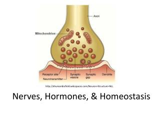
Neuron Communcation • 2. chemical • Prevalent in most other organs, including the CNS, where signaling among neurons is more complex and varied. • Have a narrow gap, called the synaptic cleft, separating a synaptic terminal of the sending neuron from the receiving neuron. • The cleft prevents the action potential in the sending neuron from spreading directly to the receiving neuron. • Instead, the action potential (electrical signal) is first converted to a chemical signal. • The chemical signal, consisting of molecules of neurotransmitter, may then generate an action potential in the receiving cell. • Neurotransmitter is contained in synaptic vesicles in the sending neuron’s synaptic terminals.
Neuron Communication • Events that occur at a chemical synapse (Figure 28.6) • 1. An action potential arrives at the synaptic terminal. • 2. The action potential triggers chemical changes that cause some synaptic vesicles to fuse with the plasma membrane of the sending cell. • 3. The fused vesicles release their neurotransmitter molecules by exocytosis into the synaptic cleft, and the neurotransmitter diffuses across the cleft. • 4. (4-6 varies among different types of chemical synapses) In one common type, the released neurotransmitter binds to receptor molecules on the receiving cell’s plasma membrane. • 5. the binding of neurotransmitter to receptor opens chemically gated ion channels in the receiving cell’s membrane • With the channels open, ions can diffuse into the receiving cell and trigger new action potentials. 6. The neurotransmitter is broken down by an enzyme or transported back into the signaling cell, and the ion channels close. *Ensures that the neurotransmitter’s effect is brief and precise.
Neuron Communication Chemical Synapse
Neuron Communication • As you read this PowerPoint… • Action potentials carrying information about the words on the SmartBoard are streaming along sensory neurons from your eyes to your brain. • Arriving at synapses with receiving cells (interneurons in the brain), the action potentials are triggering the release of neurotransmitters at the ends of sensory neurons. • The neurotransmitters are diffusing across synaptic clefts and triggering changes in some of your interneurons—changes that lead to integration of the signals and ultimately to what the signals actually mean (in this case, the meaning of words and sentences) • Next, motor neurons in your brain will send out action potentials to muscle cells in your fingers, telling them to write down this information in just the right way.
Neuron Communication • One neuron may interact with many others. • In fact, a neuron may receive information via neurotransmitters from hundreds of other neurons via thousands of synaptic terminals. • Inputs can be highly varied because each sending neuron may secrete a different quantity or kind of neurotransmitter. • If the signals are strong enough to raise the membrane potential to threshold, an action potential will be generated in the receiving cell. • That neuron then passes signals along its axon to other cells at a rate that represents a summation of all the information it has received. • ***Signal frequency is key because action potentials are all-or-none events***
Neuron Communication • What do neurotransmitters actually do to receiving neurons? • The binding of a neurotransmitter to a receptor may open ion channels in the receiving cell’s plasma membrane or trigger a signal transduction pathway that does so. • The more neurotransmitter molecules that bind to receptors on the receiving cell and the closer the synapse is to the base of the receiving cell’s axon, the stronger the effect. • For example, neurotransmitters that open Na+ channels may trigger action potentials in the receiving cell. • These neurotransmitters and synapses from which they are released are referred to as “excitatory” • In contrast, many neurotransmitters open membrane channels for ions that decrease the tendency to develop action potentials in the receiving cell—such as channels that release K+ • These neurotransmitters and synapses from which they are released are referred to as “inhibitory.”
Neurotransmitters • The propagation of nerve signals across chemical synapses depends on neurotransmitters. • Many are small, nitrogen-containing organic molecules. • Acetylcholine • Important in the brain and at synapses between motor neurons and muscle cells. • Depending on the kind of receptors on receiving cells, it may be excitatory or inhibitory. • For example, it makes our skeletal muscles contract but slows the rate of contraction of cardiac muscles.
Neurotransmitters • Biogenic amines • Derived from amino acids; nitrogen-containing. • Include epinephrine, norepinephrine, serotonin, and dopamine, all of which also function as hormones. • Important in the CNS. • Serotonin and dopamine affect sleep, mood, attention, and learning. • Imbalances of biogenic amines are associated with various disorders. • Parkinson’s disease is associated with a lack of dopamine, and an excess of dopamine is linked to schizophrenia. • Reduced levels of norepinephrine and serotonin seem to be linked with some types of depression. • Some psychoactive drugs, including LSD, apparently produce their hallucinatory effects by binding to serotonin and dopamine receptors in the brain
Neurotransmitters • Aspartate, glutamate, glycine, and GABA (gamma aminobutyric acid) • Amino acids that are known to be important in the CNS • Aspartate and glutamate are excitatory; glycine and GABA are inhibitory.
Neurotransmitters • Several peptides, relatively short chains of amino acids: • One called substance P, is an excitatory neurotransmitter that mediates our perception of pain. • Also, endorphins function as both neurotransmitters and hormones, decreasing our perception of pain during times of physical or emotional stress.
Neurotransmitters • Dissolved gases • Nitric oxide (NO); during sexual arousal in human males, certain neurons release NO into the erectile tissue of the penis, and the NO triggers an erection. • Neurons produce NO molecules on demand, rather than storing them in synaptic vesicles. • The dissolved gas diffuses into neighboring cells, produces a change, and is quickly broken down. • **The erectile dysfunction drug Viagra promotes this effect of NO**
Drugs • Drugs are used both medicinally and recreationally. • While they have the ability to increase alertness and sense of well-being or to reduce physical and emotional pain, they also have the potential to disrupt the brain’s finely tuned neural pathways, altering the chemical balances that are the product of millions of years of evolution.
Drugs • Many psychoactive drugs, even common ones such as caffeine, nicotine, and alcohol, affect the action of neurotransmitters in the brain’s billions of synapses. • Caffeine keeps us awake by countering the effects of inhibitory neurotransmitters. • Nicotine acts as a stimulant by binding to and activating acetylcholine receptors. • Alcohol is a strong depressant • Its precise effect on the nervous system is not yet known, but its seems to increase the inhibitory effects of the neurotransmitter GABA.
Drugs • Prescription drugs • Used to treat psychological disorders by altering the effects of neurotransmitters. • Most popular class of antidepressant medication works by affecting the action of serotonin. • Selective serotonin reuptake inhibitors (SSRIs), block the removal of serotonin from synapses, increasing the amount of time this mood-altering neurotransmitter is available to receiving cells. • Tranquilizers such as diazepam (Valium) and alprazolam (Xanax) activate the receptors for GABA, increasing the effect of this inhibitory neurotransmitter.
Drugs • Prescription drugs • Antipsychotic drugs are used to treat schizophrenia by blocking dopamine receptors • Drug used to treat attention deficit hyperactivity disorder (ADHD), such as methphenidate (Ritalin) are chemically similar to dopamine and norepinephrine. • ADHD medications are believed to block the reuptake of these neurotransmitters, but their precise actions are poorly understood.
Drugs • Illegal drugs • Stimulants such as amphetamines and cocaine increase the release and availability of norepinephrine and dopamine at synapses. • Abuse of these drugs can produce symptoms resembling schizophrenia. • The active ingredient in marijuana (tetrahydrocannabinol, or THC) binds to brain receptors normally used by another neurotransmitter (anandamide) that seems to play a role in pain, depression, appetite, memory, and fertility. • Opiates bind to endorphin receptors, reducing pain and producing euphoria. • Morphine, codeine, and heroin. • Can be used medicinally for pain relief • However, abuse may permenantly change the brain’s chemical synapses and reduce normal synthesis of neurotransmitters.
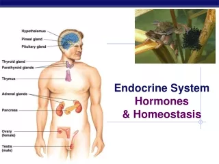
Hormones • Hormone: a chemical signal that is carried by the circulatory system (usually in the blood) and that communicates regulatory messages within the body. • Endocrine glands are the organs that make and secrete hormones. • Endocrine system contain all of an animal’s hormone-secreting cells and serves as the body’s main chemical-regulating system. • Because most hormones are carried in the blood, they reach all parts of the body, and the endocrine system is thus especially important in controlling whole-body activities. • For example, hormones coordinate responses to stimuli such as stress, dehydration, or low blood glucose levels. • Hormones also regulate long-term developmental processes, such as growth, maturation, and reproduction.
Hormones • Hormones may travel to all parts of the body, but only certain types of cells, called target cells, are equipped to respond. • A single hormone molecule may dramatically alter a target cell’s metabolism by turning on or off the production of a number of enzymes. • A tiny amount of a hormone can govern the activities of enormous numbers of target cells in a variety of organs. • A hormone is ignored by other types of cells (nontarget cells)