PLACENTA
PLACENTA. Placenta (plah-sen’tah) is a Latin word. It means “a flat cake”. Placenta is an organ characteristic of mammals, developing during pregnancy, joining mother and offspring, providing necessary provisions for the sustenance of developing human in intrauterine life.


PLACENTA
E N D
Presentation Transcript
Placenta (plah-sen’tah) is a Latin word. It means “a flat cake”. • Placenta is an organ characteristic of mammals, developing during pregnancy, joining mother and offspring, providing necessary provisions for the sustenance of developing human in intrauterine life.
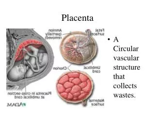
The placenta is an organ that connects the developing fetus to the uterine wall to allow nutrient uptake, waste elimination, and gas exchange via the mother's blood supply. • "True" placentas are a defining characteristic of eutherian or "placental" mammals,
Eutheria (/juːˈθɪəriə/; from Ancient Greek ευθήριον, euthērion, meaning "true/good beasts") is the clade consisting of primates and all other mammals—in many orders—that are more closely related to them than they are to marsupials.
Life restoration of Juramaia sinensis Kingdom Animalia Phylum Chorda Class Mammalia Branch Eutheria
Marsupials are an infraclass of mammals living primarily in the Southern Hemisphere; a distinctive characteristic, common to most species, is that the young are carried in a pouch.
Female Eastern Grey Kangaroo with a joey in her pouch Kingdom Animalia Phylum Chorda Class Mammalia Infraclass Marsupials
A clade (from Ancient Greek κλάδος, klados, "branch") or monophylum (see monophyletic) is a group consisting of an ancestor and all its descendants, a single "branch" on the "tree of life"
The fully developed placenta is discoid-shape with a diameter of 15 to 25 cm and is approximately 3 cm thick. • It weighs about 500 to 600 gm (about one-sixth that of the fetus).
In humans, the placenta averages 22 cm (9 inch) in length and 2–2.5 cm (0.8–1 inch) in thickness, with the center being the thickest, and the edges being the thinnest. • It typically weighs approximately 500 grams (1 lb).
It has a dark reddish-blue or crimson color. • It connects to the fetus by an umbilical cord of approximately 55–60 cm (22–24 inch) in length, which contains two umbilical arteries and one umbilical vein
The umbilical cord inserts into the chorionic plate (has an eccentric attachment). • Vessels branch out over the surface of the placenta and further divide to form a network covered by a thin layer of cells. This results in the formation of villous tree structures.
On the maternal side, these villous tree structures are grouped into lobules called cotyledons. • In humans, the placenta usually has a disc shape, but size varies vastly between different mammalian species
Placenta covers 15 to 30 % of the decidua (endometrium during pregnancy)
Placenta is a fetomaternal organ that has two components: • A fetal portion that develops from a portion of chorionic sac, called chorionic frondosum. • A maternal portion that is derived from a portion of endometrium, called decidua basalis.
The placenta begins to develop upon implantation of the blastocyst into the maternal endometrium. • Placenta grows throughout pregnancy. • Development of the maternal blood supply to the placenta is complete by the end of the first trimester of pregnancy (approximately 12–13 weeks).
Fertilization - Zygote The cleavage starts in the zygote immediately after fertilization and on 4th day morula has formed. The morula consists of two groups of cells: Inner Cell Mass (Central Cells) Outer Cell Mass (Peripheral Cells)
Within one day morula is converted into blastocyst consisting of same two groups of cells, now with different names: Embryoblast derived from Inner Cell Mass Trophoblast derived from Outer Cell Mass
Embryoblast forms the embryo proper • Trophoblast forms the placenta and associated membranes.
Development of placenta starts as soon as blastocyst is attached to the endometrium.
Trophoblasts start proliferating rapidly and differentiate into two layers: • Cytotrophoblast or cellular trophoblast • Syncytial trophoblast (syncytiotrophoblast)
Transverse section of Secondary Villous (day 16)
Meanwhile, the cytotrophoblastic cells in the villi penetrate progressively into the overlying syncytial trophoblast until they reach maternal endometrium. Here they establish contact with similar extensions of neighboring villous stems, thus forming a thin outer cytotrophoblast shell. This shell gradually surrounds the trophoblast entirely. Cytotrophoblastic shell attaches chorionic sac to endometrial tissue.
Stem Villi Villi that are attached to the maternal tissues via cytotrophoblastic shell are called stem villi or anchoring villi. Floating Villi The villi that branch from stem villi and float free in intervillous space are called branching villi or floating villi. These villi are not attached to maternal tissue.
In the villi, these vessels eventually branch to form an extensive arterio-capillary-venous system, bringing the fetal blood extremely close to the maternal blood; but no intermingling of fetal and maternal blood occurs ("placental barrier")
Tertiary villous of 10 weeks old Fetus • Blood Placental Barrier is formed by: • Syncytial trophoblast • Cytotrophoblast • Mesoderm or Connective tissue • Endothelium of fetal blood vessels
Tertiary villous of full term Fetus. • Blood Placental Barrier is formed by: • Syncytial trophoblast • Endothelium of fetal blood vessels
In preparation for implantation, the uterine endometrium undergoes "decidualisation". • Spiral arteries in decidua are remodeled so that they become less convoluted and their diameter is increased.
The increased diameter and straighter flow path both act to increase maternal blood flow to the placenta. • The relatively high pressure as the maternal blood fills intervillous space through these spiral arteries bathes the fetal villi in blood, allowing an exchange of gases to take place.
In humans and other hemochorial placentals, the maternal blood comes into direct contact with the fetal chorion, though no fluid is exchanged. • As the pressure decreases between pulses, the deoxygenated blood flows back through the endometrial veins. • Maternal blood flow is approximately 600–700 ml/min at term.
Schematic diagram showing the direction of maternal flow through cotyledons.