Download

1 / 29
370 likes | 1.95k Vues
CAPILLARY ELECTROPHORESIS. Prepared by Dr : Sherif A.Abdel-Gawad. Electrophoresis.

E N D
CAPILLARY ELECTROPHORESIS Prepared by Dr: SherifA.Abdel-Gawad
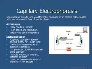
Electrophoresis • It is another class of separation techniques in which analytes are separated based on their ability to move through a conductive medium, usually an aqueous buffer, in response to an applied electric field. • In capillary electrophoresis the conducting buffer is retained within a capillary tube whose inner diameter is typically 25–75 mm. • Samples are injected into one end of the capillary tube. Under the effect of applied electric field, the sample migrates through the capillary, its components separate and elute from the column at different times. • The resulting electropherogramlooks similar to the chromatograms obtained in GC or HPLC and provides both qualitative and quantitative information.
Theory of Capillary Electrophoresis • In capillary electrophoresis the sample is injected into a buffered solution retained within a capillary tube. When an electric field is applied to the capillary tube, the sample’s components migrate as the result of two types of mobility: electrophoretic mobility and electroosmotic mobility. • Electrophoretic mobilityis the solute’s response to the applied electric field. Cations move toward the negatively charged cathode, anions move toward the positively charged anode, and neutral species, which do not respond to the electric field, remain stationary. • The other contribution to a solute’s migration is electroosmotic flow, which occurs when the buffer solution moves through the capillary in response to the applied electric field. • Under normal conditions the buffer solution moves toward the cathode, sweeping most solutes, even anions, toward the negatively charged cathode.
Electrophoretic Mobility • The velocity with which a solute moves in response to the applied electric field is called its electrophoretic velocity, it is defined as Electrophoretic velocity = µE where µis the solute’s electrophoretic mobility, and E is the magnitude of the applied electric field. • A solute’s electrophoretic mobility is defined as µ = q/6ɳπr where q is the solute’s charge, ɳis the buffer solvent’s viscosity, and r is the solute’s radius. • From the above equations, electrophoretic mobility, and, therefore, electrophoretic velocity, is largest for more highly charged solutes and solutes of smaller size. Since q is positive for cations and negative for anions, these species migrate in opposite directions. Neutral species, for which q is 0, have an electrophoretic velocity of 0.
Electroosmotic Mobility • What is observed under normal conditions, however, is that the buffer solution moves toward the cathode. This phenomenon is called the electroosmoticflow. • Electroosmosis occurs because the walls of the capillary tubing are electrically charged. The surface of a silica capillary contains large numbers of silanolgroups (Si–OH). At pH levels greater than approximately 2 or 3, the silanol groups ionize to form negatively charged silanate ions (Si–O–). Cations from the buffer are attracted to the silanate ions.
From the above figure, some of the cationsbind tightly to the silanate ions, forming an inner, or fixed, layer. Other cations are more loosely bound, forming an outer, or mobile, layer. Together these two layers are called the double layer. • Cations in the outer layer migrate toward the cathode. Because these cations are solvated, the solution is also pulled along, producing the electroosmoticflow.
Electroosmotic flow velocity is a function of the magnitude of the applied electric field and the buffer solution’s electroosmoticmobility (µeof) Electroosmotic flow velocity = µeofE Electroosmotic mobility is defined as µeof = ɳπ where is the buffer solution’s dielectric constant, is the zeta potential, and ɳ is the buffer solution’s viscosity. • From the above equations, zeta potential plays an important role in determining the electroosmotic flow velocity. The zeta potential is directly proportional to the charge on the capillary walls, with a greater density of silanate ions corresponding to a larger zeta potential. • Below a pH of 2, for example, there are few silanate ions; thus, the zeta potential and electroosmoticflow velocity are 0. As the pH level is increased, both the zeta potential and the electroosmotic flow velocity increase.
N.B. The electroosmotic flow profile is very different from that for a phase moving under forced pressure. The flow profile for electroosmosiswith that for hydrodynamic pressure. The uniform, flat profile for electroosmosis helps to minimize band broadening in capillary electrophoresis, thus improving separation efficiency.
Total Mobility • A solute’s net, or total velocity, is the sum of its electrophoretic velocity and the electroosmotic flow velocity. • Under normal conditions the following relationships hold: (vtot)cations> veof (vtot)anions < veof (vtot)neutrals = veof • Thus, cations elute first in an order corresponding to their electrophoretic mobilities, with small, highly charged cations eluting before larger cations of lower charge. Neutral species elute as a single band, with an elution rate corresponding to the electroosmoticflow velocity. Finally, anions are the last components to elute, with smaller, highly charged anions having the longest elution time.
N.B. • The solute migration time is governed by the following relation: Where l is the distance from the site of injection to the detector site, L is the length of the capillary tube, V is the applied potential. • So we can decrease a solute’s migration time (and thus the total analysis time) by applying a higher voltage or by using a shorter capillary tube. Increasing the electroosmotic flow also shortens the analysis time, but, as we will see shortly, at the expense of resolution.
Efficiency • The efficiency of capillary electrophoresis is characterized by the number of theoretical plates, N, just as it is in GC or HPLC. • In capillary electrophoresis, the number of theoretic plates is determined by the following equation: where D is the solute’s diffusion coefficient. • From the above equation it is easy to see that the efficiency of a capillary electrophoretic separation increases with higher voltages. • Again, increasing the electroosmotic flow velocity improves efficiency, but at the expense of resolution. • Efficiency in capillary electrophoresis is independent on the capillary’s length (opposite to chromatography). • Typical theoretical plate counts are approximately 100,000–200,000 for capillary electrophoresis.
Selectivity • In chromatography, selectivity is defined as the ratio of the capacity factors for two solutes. In capillary electrophoresis, the analogous expression for selectivity is where and are the electrophoretic mobilities for solutes 1 and 2, respectively, chosen such that α>1. • Selectivity often can be improved by adjusting the pH of the buffer solution. • For example, ammonium ion is a weak acid with a pKa of 9.24. At a pH of 9.24 the concentrations of ammonium ionand ammonia neutral molecule are equal. Decreasing the pH below 9.24 increases its electrophoretic mobility because a greater fraction of the solute is present as the cation. On the other hand, raising the pH above 9.24 increases the proportion of the neutral ammonia, decreasing its electrophoretic mobility.
Resolution • The resolution between two solutes is where av is the average electrophoretic mobility for the two solutes. • From the above equation, increasing the applied voltage and decreasing the electroosmotic flow velocity improves resolution. • The latter effect is particularly important because increasing electroosmotic flow improves analysis time and efficiency while decreasing resolution.
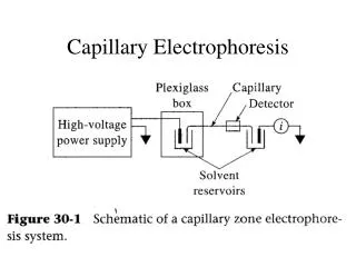
INSTRUMENTATION The basic instrumentation for capillary electrophoresis includes a power supply for applying the electric field, anode and cathode compartments containing reservoirs of the buffer solution, a sample vial containing the sample, the capillary tube, and a detector.
Capillary Tubes • Most capillary tubes are made from fused silica coated with a 20–35-µm layer of polyimide to give it mechanical strength. The inner diameter is typically 25–75 µm, which is smaller than that for a capillary GC column, with an outer diameter of 200–375 µm. • The narrow bore of the capillary column and the relative thickness of the capillary’s walls are important. When an electric field is applied to a capillary containing a conductive medium, such as a buffer solution, current flows through the capillary.
This current leads to Joule heating, the extent of which is proportional to the capillary’s radius and the magnitude of the electric field. • Joule heating is a problem because it changes the buffer solution’s viscosity, with the solution at the center of the capillary being less viscous than that near the capillary walls. Since the solute’s electrophoretic mobility depends on the buffer’s viscosity and so solutes in the center of the capillary migrate at a faster rate than solutes near the capillary walls. . • The result is additional band broadening that degrades the separation. • Capillaries with smaller inner diameters generate less Joule heating, and those with larger outer diameters are more effective at dissipating the heat.
Injecting the Sample • The mechanism by which samples are introduced in capillary electrophoresis is quite different from that used in GC or HPLC. Two types of injection are commonly used: hydrodynamic injection and electrokinetic injection. • In both cases the capillary tube is filled with buffer solution. One end of the capillary tube is placed in the destination reservoir, and the other is placed in the sample vial. Hydrodynamic injection • It uses pressure to force a small portion of the sample into the capillary tubing. • To inject a sample hydrodynamically a difference in pressure is applied across the capillary by either pressurizing the sample vial or by applying a vacuum to the destination reservoir.
Electrokineticinjection • It is made by placing both the capillary and the anode into the sample vial and briefly applying an electric field so the introduction of the sample is mainly dependent on the electrophoretic mobility of the samples. • The solutes with the largest electrophoretic mobilities (smaller, more positively charged ions) are injected in greater numbers than those with the smallest electrophoretic mobilities (smaller, more negatively charged ions).
Stacking • When a solute’s concentration in the sample is very small, it may be possible to inject the solute in a manner that increases its concentration in the capillary tube. This method of injection is called stacking. • Stacking is accomplished by placing the sample in a solution whose ionic strength is significantly less than that of the buffering solution. Because the sample plug has a lower concentration of ions than the buffering solution, its resistance is greater. Since the electric current passing through the capillary is fixed, so the electric field in the sample plug is greater than that in the buffering solution. Ohm Law E = iR
Since electrophoretic velocity is directly proportional to the electric field ,thus, ions in the sample plug migrate with a greater velocity. When the solutes reach the boundary between the sample plug and the buffering solution, the electric field decreases and their electrophoretic velocity slows down, “stacking” together in a smaller sampling zone as shown in the following figure
Applying the Electric Field • Migration in electrophoresis occurs in response to the applied electric field. • Application of large electric field is important because higher voltages lead to shorter analysis times, more efficient separations, and better resolution. • Voltages up to 40 kV can be applied.
Detectors • Most of the detectors used in HPLC also find use in capillary electrophoresis. • UV/Vis detectors are among the most popular. Because absorbance is directly proportional to path length, the capillary tubing’s small diameter leads to signals that are smaller than those obtained in HPLC. • Several approaches have been used to increase the path length, including a Z-shaped sample cell and bubble cell. • Better detection limits are obtained using fluorescence, particularly when using a laser as an excitation source.
Capillary Electrophoresis Methods 1- Capillary Zone Electrophoresis (CZE). • In CZE the capillary tube is filled with a buffer solution and, after loading the sample, the ends of the capillary tube are placed in reservoirs containing additional buffer solution. • Under normal conditions, the end of the capillary containing the sample is the anode, and solutes migrate toward the cathode at a velocity determined by their electrophoretic mobility and the electroosmotic flow. • Cationselute first, with smaller, more highly charged cationseluting before larger cations with smaller charges. • Neutral species elute as a single band. Finally, anions are the last species to elute, with smaller, more negatively charged anions being the last to elute.
The direction of electroosmotic flow and, therefore, the order of elution in CZE can be reversed. This is accomplished by adding an alkylammonium salt to the buffer solution. The positively charged end of the alkylammonium ion binds to the negatively charged silanate ions on the capillary’s walls. • The alkylammonium ion’s “tail” is hydrophobic and associates with the tail of another alkylammonium ion. • The result is a layer of positive charges to which anions in the buffer solution are attracted. • The migration of these solvated anions toward the anode reverses the electroosmotic flow’s direction. The order of elution in this case is exactly the opposite of that observed under normal conditions.
Capillary zone electrophoresis also can be accomplished without an electroosmotic flow by coating the capillary’s walls with a nonionic reagent. • In the absence of electroosmoticflow only cations migrate from the anode to the cathode. • Capillary zone electrophoresis provides an effective separations of any charged species, including inorganic anions and cations, organic acids and amines, and large biomolecules such as proteins. For example, CZE has been used to separate a mixture of 36 inorganic and organic ions in less than 3 minutes. Neutral species, of course, cannot be separated.
2- MicellarElectrokinetic Capillary Chromatography • One limitation to CZE is its inability to separate neutral species. Micellarelectrokineticchromatography (MEKC) overcomes this limitation by adding a surfactant, such as sodium dodecylsulfate to the buffer solution. • Sodium dodecylsulfate, (SDS) has a long-chain hydrophobic “tail” and an ionic functional group, providing a negatively charged “head.” When the concentration of SDS is sufficiently large, a micelle forms. A micelle consists of an agglomeration of 40–100 surfactant molecules in which the hydrocarbon tails point inward, and the negatively charged heads point outward. • Because micelles are negatively charged, they migrate toward the cathode with a velocity less than the electroosmotic flow velocity.
Neutral species partition themselves between the micelles and the buffer solution in much the same manner as they do in HPLC. Because there is a partitioning between two phases, the term “chromatography” is used. • Note that in MEKC both phases are “mobile.” The elution order for neutral species in MEKC depends on the extent to which they partition into the micelles. • Hydrophilic neutrals are insoluble in the micelle’s hydrophobic inner environment and elute as a single band as they would in CZE. Neutral solutes that are extremely hydrophobic are completely soluble in the micelle, eluting with the micelles as a single band. Those neutral species that exist in a partition equilibrium between the buffer solution and the micelles elute between the completely hydrophilic and completely hydrophobic neutrals. • Micellarelectrokineticchromatography has been used to separate a wide variety of samples, including mixtures of pharmaceutical compounds, vitamins, and explosives.
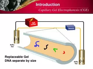
3- Capillary Gel Electrophoresis • In capillary gel electrophoresis (CGE) the capillary tubing is filled with a polymeric gel. Because the gel is porous, solutes migrate through the gel with a velocity determined both by their electrophoretic mobility and their size. • The ability to effect a separation based on size is useful when the solutes have similar electrophoretic mobilities. For example, fragments of DNA of varying length have similar charge-to-size ratios, making their separation by CZE difficult. Since the DNA fragments are of different size, a CGE separation is possible. • The capillary used for CGE is usually treated to eliminate electroosmoticflow, thus preventing the gel’s extrusion from the capillary tubing. • Samples are injected electrokineticallybecause the gel provides too much resistance for hydrodynamic sampling. • The primary application of CGE is the separation of large biomolecules, including DNA fragments, proteins, and oligonucleotides.
4- Capillary Electrochromatography • Another approach to separating neutral species is capillary electrochromatography(CEC). • In this technique the capillary tubing is packed with 1.5–3-mm silica particles coated with a bonded, nonpolar stationary phase. • Neutral species separate based on their ability to partition between the stationary phase and the buffer solution (the mobile phase). • Separations are similar to the analogous HPLC separation, but without the need for high-pressure pumps. • The efficiency in CEC is better than in HPLC, with shorter analysis times.