Morphological Features and Diagnosis of Transplant Glomerulitis: A Case Study
This case study presents the morphological features observed in a 39-year-old male following renal transplantation. Key findings include increased endocapillary leukocytes, endothelial swelling, dilatation of capillary loops, and occlusion of capillary lumina. The differential diagnosis highlights transplant glomerulitis and endocapillary glomerulonephritis. The case emphasizes the importance of cellular composition, including monocytes and lymphocytes, and their roles in the pathogenesis of renal rejection. Follow-up reveals significant complications, underscoring the critical nature of careful monitoring post-transplantation.

Morphological Features and Diagnosis of Transplant Glomerulitis: A Case Study
E N D
Presentation Transcript
Case 6 Helmut Hopfer, University Hospital Basel, Switzerland
morphologicalfeatures • increasednumberofendocapillaryleukocytes • endothelialswelling • dilatationofcapillaryloops • occlusionornearocclusionofcapillaryluminawithcells usually a focaland segmental lesion!
EM differential diagnosis • transplant glomerulitis • endocapillaryglomerulonephritis • (intravascularlymphoma)
transplant glomerulitis: definition • recognizing a pattern: • increasednumberofendocapillaryleukocytes • endothelialswelling • dilatationofcapillaryloops • occlusionornearocclusionofcapillaryluminawithcells • countingmononuclearcells (arbitrarycut-off) → nocleardistinctionbetweencelltypesby light microscopy! → immunohistochemistry, definitionofcut-off by ROC Example: endocapillary immune-complex GN CD20 CD68 CD5 ERG
2. 1. 3.
discussion & conclusions • Whyaretheleukocytesthere? • Whatarethemonocytes / macrophagesdoingthere? • Whatarethelymphocytesdoingthere? → monocytesexert an earlyendocapillaryreparativefunction on theendothelialcells
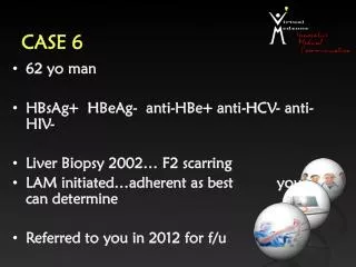
casepresentation Clinical history: 39 yearold male. Renal transplantation (TR) in 2005 due tohypertension. Malcompliancewithimmunosuppresion → twoepisodesofinterstitialcellularrejection. Diagnosticbiopsy (BX) 40 months after TR, rise in creatinineandnewlydiagnosedproteinuria. Diagnosis: Transplant glomerulitis (by EM), severe diffuse interstitialcellularrejection, C4d negative. Focal IFTA (10-20%). Follow up: Dialysisdependence 7 monthslater. BX withmixed T cell- and antibody-mediatedrejection, C4d positive.