The Journey from Fertilization to Birth: A Comprehensive Overview of Human Development
420 likes | 534 Vues
This chapter delves into the extraordinary journey of human development from fertilization to birth. It explores the critical events post-fertilization, highlighting the union of sperm and egg, the stages of fertilization, cleavage, and blastocyst formation. The process of implantation, placentation, and embryonic development is examined, detailing the formation of extra-embryonic membranes and germ layers. It further elaborates on organogenesis, fetal development, and the key milestones that occur between conception and the critical stages of prenatal growth.

The Journey from Fertilization to Birth: A Comprehensive Overview of Human Development
E N D
Presentation Transcript
Biology 221 Anatomy & Physiology II TOPIC 13Survey of Development Chapter 29 and others E. Lathrop-Davis / E. Gorski / S. Kabrhel
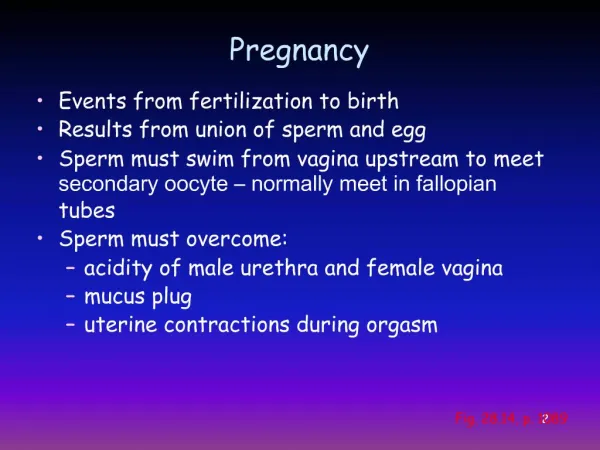
Pregnancy • Events from fertilization to birth • Results from union of sperm and egg • Sperm must swim from vagina upstream to meet secondary oocyte – normally meet in fallopian tubes • Sperm must overcome: • acidity of male urethra and female vagina • mucus plug • uterine contractions during orgasm Fig. 28.14, p. 1089
Fertilization • union of haploid gametes (egg & sperm) produces diploid zygote • occurs in 2 stages: • Stage 1: penetration of egg • Stage 2: union of sperm & egg membranes and nuclei
Fertilization: Stage 1 - Penetration • Acrosomal enzymes from many, many sperm must be released to break through corona radiata and zona pellucida • When one sperm finally makes it through, conditions in the oocyte change so that no more sperm can enter Fig. 29.2, p. 1121
Fertilization: Stage 2 Union • Oocyte undergoes meiosis II and ejects the 2nd polar body • Sperm nucleus migrates to center of oocyte, rest of sperm degenerates • Sperm and egg nuclei unite to form diploid zygote Fig. 29.3, p. 1122
Characteristics of Living Things • Maintenance of boundaries (cell membrane) • Movement (cellular and organismal) • Respond to stimuli (recognize changes in environment and creates responses) • Digest (provides nutrients in usable form) • Metabolism (chemical reactions) • Excretion (removal of wastes) • Reproduction (cellular) • Growth (increase in size)
Pre-embryonic Development1st Through 2nd Weeks Overview • Cleavage • Blastocyst foramtion • Implantation • Placentation
Cleavage &Blastocyst Formation • Cleavage – rapid replication of DNA and mitotic cell divisions produce ever smaller cells resulting in a solid ball called a morula • Continued division produces blastocyst • inner cell mass becomes future embryo • trophoblast cells form fetal part of placenta Fig. 29.4, p. 1123
Implantation • blastocyst implants itself into endometrium of uterine wall • ectopic pregnancy – implantation occurs in some other location (e.g., fallopian tube) • trophoblast cells (outer cells of blastocyst) secrete human chorionic gonadotropin (hCG) that maintains corpus luteum through 1st four months Fig. 29.5, p. 1124
Placentation • development of placenta • originates from chorion (trophoblast cells) of embryo/fetus and endometrial tissue of mother • begins to produce estrogens and progestins Fig. 29.7a-c, p. 1126
Embryonic DevelopmentWeeks 3-8 Overview • Development of extra-embryonic membranes • Gastrulation (formation of primary germ layers) • Organogenesis (formation of rudiments of organ systems)
Development of Extra-Embryonic Membranes • chorion – forms part of placenta • anmion • becomes filled with amniotic fluid: • cushions embryo • maintains temperature • allows freedom of movement • yolk sac • forms part of the primitive gut • 1st site of blood cell formation • allantois • contributes to the umbilical cord • becomes part of urinary bladder Fig. 29.7d, p. 1126
Gastrulation • development of Primary Germ Layers • ectoderm – outmost – forms epidermis and nervous system • endoderm – inner layer – forms epithelial linings of digestive tract, respiratory tract, urogenital system and associated glands • mesoderm – middle layer – forms connective tissues and muscle See also Table 29.1, p. 1135 Fig. 29.8, p. 1131
Specialization of Ectoderm • over most of body, forms epidermis • over middle back, forms neural plate neural groove & neural folds neural tube brain & spinal cord Fig. 29.9, p. 1132
Specialization of Endoderm • gives rise to lining of gut • structures that come from gut arise as “outpocketings” • lungs and respiratory tree (epithelial linings) • thyroid, parathyroid glands • liver and gall bladder • pancreas Fig. 29.10, p. 1133
Specialization of Mesoderm • gives rise to muscle, connective tissues, serous membranes • limb buds form and move laterally in what will become shoulder and hip areas Fig. 29.11, p. 1134
Embryonic developmentWeeks 3-8: Organogenesis • heart beats by week 4 • all systems present in some form by week 8 • all major regions of brain present by week 8 • liver produces blood cells by week 8
Fetal Development: 12 - 16 Weeks By 12 weeks: • blood cell formation in bone marrow • ossification begins By 16 weeks: • kidneys have typical shape • joint (synovial) cavities present • cerebellum becomes large • sensory organs differentiated See Table 29.2, p. 1138
Fetal Development: 20-30 Weeks • by 20 weeks • skin covered by lanugo (silky hair) • activity can be felt by mother (“quickening”) • by 30 weeks • myelination of spinal cord begins • finger and toe nails present • bone marrow becomes only site of blood cell formation • testes descend (7th month) in males • surfactant production begins at ~ 24 weeks See Table 29.2, p. 1138
Fetal Development:8 Months - Birth • 8th to 9th months • continued development of organ systems • significant weight gain • weeks 38-42 – birth • before 38 weeks, less fat, organ systems not as well developed • after 42 weeks, placenta begins to degenerate See Table 29.2, p. 1138
Development of Integumentary System (Ch. 5) • Pp. 165-168 • epidermis and dermis developed by 4th month • epidermal derivatives grow down into dermis • lanugo present from 20 weeks • vellus hairs present by 7th month http://www.uoguelph.ca/zoology/devobio/210labs/ecto5.html
Development of Skeletal System • Ch. 6; P. 181 • begins by 8th week • primary ossification completed by birth • secondary ossification continues to early adulthood http://www.uoguelph.ca/zoology/devobio/210labs/meso2.html#osteo
Development of Skeletal System • endochondral ossification – in hyaline cartilage models of most bones other than cranial bones and clavicles • intramembranous ossification – in flat bones of cranium and clavicles http://www.uoguelph.ca/zoology/devobio/210labs/meso2.html#osteo
Development of Skeletal System • fontanels – unossified membranes in skull at birth; allow head to change shape slightly for easier birth http://www.bio.psu.edu/faculty/strauss/anatomy/skel/fetal.htm
Development of Spinal Curvatures • primary curvatures – thoracic and sacral – present at birth • secondary curvatures – cervical and lumbar – develop as infant lifts head and stands, respectively http://www.csu.edu.au/faculty/arts/humss/bioethic/abort1.htm
Development of Nervous System • Ch. 11; pp. 429-430, 463-464 • develops from “neural ectoderm” • neural crest cells (adjacent to tube) give rise to sensory neurons • neural tube cells give rise to interneurons and motor neurons http://www.angelfire.com/mb/jessicasjourney/info.html Fig. 12.2, p. 430
Development of Nervous System • eyes develop as outgrowth of diencephalon • brain and spinal cord develop from neural tube • brain regions represent enlargements of anterior tube • ventricles develop from openings in neural tube • anencephaly – failure of cerebrum and part of brain stem to develop Fig. 12.4, p. 431
Development of Nervous System • spinal cord develops from middle and posterior portions of tube • spina bifida • incomplete fusion of vertebral arches, usually in lumbrosacral region • up to 70% of cases associated with inadequate folate levels in mother • some cases associated with mother’s exposure to high levels of UV radiation [DISCOVER Vol. 22 No. 2 (February 2001)] http://www.abbottdiagnostics.com/medical_conditions/fertility_pregnancy/afp.htm
Development of Endocrine System • complex development including all three germ layers • two glands in particular develop from two different layers • pituitary • adrenal http://anatomy.med.unsw.edu.au/cbl/embryo/Notes/endocrine9.htm
Development of Pituitary Gland • adenohypophysis (anterior) develops from endoderm (roof of primitive mouth) • neurohypophysis (posterior) develops from neural ectoderm as extension of diencephalon (hypothalamus) http://www.teaching-biomed.man.ac.uk/histology/T270.HTML See also http://anatomy.med.unsw.edu.au/cbl/embryo/Notes/endocrine7.htm
Development of Adrenal Gland • cortex develops from mesoderm • medulla develops from neural ectoderm http://sprojects.mmi.mcgill.ca/embryology/ug/Adrenal_Stuff/Normal/zones.html
Development of Circulatory System • Blood – bone marrow, yolk sac • Heart – thoracic cavity • Blood vessels – start in yolk sac
Development of Blood • develops in yolk sac, liver, spleen, bone marrow of fetus • fetal hemoglobin (HbF) has a greater affinity for O2 than adult hemoglobin does http://www.lab.anhb.uwa.edu.au/mb140/CorePages/Blood/Images/bma10he.jpg http://www.smbs.buffalo.edu/bch/faculty/garrett_sinha.html
Development of Heart • begins as 2 tubes that fuse by 4th week • begins pumping in 1st month (4th week) • foramen ovale allows blood to flow from right to left atrium • moves oxygenated blood more quickly into general circulation • partially by-passes developing lungs Fig. 19.24, p. 709
Fetal Circulation • umbilical arteries – carry partially oxygenated blood to placenta • umbilical vein – returns oxygenated blood from placenta to fetal liver • ductus venosus – shunt through liver connecting umbilical vein to inferior vena cava • ductus arteriosus – connects pulmonary trunk to aorta Fig. 29.13, p. 1136
Development of Respiratory System • develops as buds from throat • surfactant production begins in week 24 • not produced in sufficient quantities until about weeks 32-35 • infant respiratory distress syndrome Fig. 23.28, p. 877
Development of Digestive System • epithelium develops from endoderm; muscle develops from mesoderm • glands develop as buds from tube Fig. 24.37, p. 938
Development of Urinary System • kidneys development begins in 4th week, completed by week 9 Fig. 26.21, p. 1035
Development of Reproductive System • ovaries & testes develop in abdominal cavity • first 6 weeks embryonic reproductive organs are “bipotential” • differentiation begins during week 7-8 under influence of testosterone • testes descend into scrotum during 7th month Fig. 28.24, p. 1106; Fig. 28.25, p. 1108
Parturition (Birth): Stages of Labor • dilation stage – cervix dilates to ~ 10 cm (4”) • expulsion stage – delivery of fetus • placental stage – delivery of placenta Fig. 29.17, p. 1142
Hormonal Control of Labor • Estrogen from ovaries induces myometrium to make more oxytocin receptors • Oxytocin from posterior pituitary (made by hypothalamus) stimulates: • Contraction of uterus • Production of prostaglandins • Stimulates contraction of uterus • Stimulates oxytocin secretion • Positive feedback cycle continues until delivery of placenta (breast feeding causes stimulation of oxytocin secretion and helps return uterus to prepregnancy size) Fig. 29.16, p. 1141
Hormonal Control of Lactation • Stimulation of pressure receptors (pressoceptors) in breast sends sensory impulses to hypothalamus • Hypothalamus stimulates posterior pituitary to release oxytocin • Oxytocin stimulates release of milk from breast • Baby continues sucking until he/she is full • After baby stops sucking, hypothalamus is no longer stimulated. Fig. 29.18, p. 1144