Logo
White Natural. Natural plastic. White plastic. 5 l 10l 50l water. (Amount of Fluorescein per well). The Effect of White Pigmentation in 96-well Plates During QPCR. Saima N. Nayab 1 , Philip N. Harries 2 , Simon C. Baker 2 and Ian Kavanagh 1†

Logo
E N D
Presentation Transcript
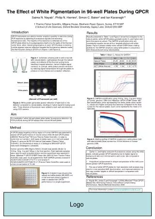
White Natural Natural plastic White plastic 5l 10l 50l water (Amount of Fluorescein per well) The Effect of White Pigmentation in 96-well Plates During QPCR Saima N. Nayab1, Philip N. Harries2, Simon C. Baker2 and Ian Kavanagh1† 1 Thermo Fisher Scientific, ABgene House, Blenheim Road, Epsom, Surrey, KT19 9AP 2 School of Life Sciences, Oxford Brookes University, Gypsy Lane, Oxford OX3 0BP Introduction Results QPCR instruments are able to monitor amplicon quantity in real-time during PCR reactions by detecting fluorescence signals and recording fluorescence data. The majority of fluorescence is reflected out of the QPCR plate either by the polypropylene itself or by the walls of the thermal cycler block, when natural polypropylene is used. QPCR plates containing a white pigment improve reflection towards the fluorescence detector inside the thermocycler during a QPCR run (Figure 1 and Figure 2). Results presented in Table 1 and Figure 3 show that compared to the natural plates, white QPCR plates generate earlier CT and higher end-point fluorescence values. The GAPDH amplification plots are consistently superior across all four template dilutions with white plates. Figure 4 shows notably more refined SYBRGreen melting profiles for the GAPDH amplicon using white plates in comparison with those produced using natural plates. • Figure 1. Schematic showing well-to-well cross talk with natural plastic. Light passes through the natural plastic and reflects off the thermal cycling block, resulting in signal refraction, hence a higher signal variance. In contrast, white plates prevent refraction by reducing signal transmission through the plastic to produce a more intense and consistent reflection. Figure 3:GAPDH amplification using 100ng, 10ng, 1ng and 100pg of human genomic DNA and ABsolute QPCR SYBR Green Mix. Red amplification plots representing the white plates show earlier CT values and higher end-point fluorescence compared to the blue curves for the natural plates. Each curve represents the mean of 8 replicates. Figure 2: White plastic generates greater reflection of light back to the detector compared to natural plates, resulting in higher signal-to-background ratio. Three dilutions of fluorescein were added to each well and detected using CCD camera. Aim To investigate if white 96-well plates allow better fluorescence detection of PCR products during QPCR assays than natural 96-well plates. Method A QPCR assay targeting a 94 bp region of human GAPDH was selected to assess QPCR performance of natural versus white 96-well QPCR plates (AB0600 Thermo-Fast ® 96 from Thermo Fisher Scientific). In order to avoid inter-run variation, both types of plates were cut in half and sealed (Figure 3) using an ALPS 50V manual heat sealer (Thermo Fisher Scientific), for simultaneous assay in a Stratagene MX3005P QPCR instrument (Stratagene, Leicester). Human genomic DNA (Sigma Aldrich, Dorset) was serially diluted to 100ng, 10ng, 1ng and 100pg in the final reaction. Eight replicate samples were used for each template dilution and the experiment was repeated 3 times. ABsolute™ QPCR SYBR® Green Mix (AB1162 from Thermo Fisher Scientific) was used for all experiments. Both GAPDH primers (forward 5' ACAGTCAGCCGCATCTTCTT 3' and reverse 5'ACGACCAAATCCGTTGACTC 3' (Thermo Fisher Scientific) were used at a final concentration of 70 nM. Figure 4: Melting profiles of GAPDH amplicons in white plates (red) and natural plates (blue) across four 10-fold dilutions of human genomic DNA. Conclusion • Earlier CT and higher end-point fluorescence values using the white plates, indicate greater QPCR sensitivity due to higher signal-to-background ratio and minimal well-to-well cross- talk compared to natural plates. • A significant enhancement in shape and precision of the melt curve shows greater QPCR specificity. • The use of white QPCR plates for high resolution melt analysis may result in more sensitive SNP discrimination, and improved detection of low copy number targets or difficult templates in comparison with natural plates. • Figure 3: A digital image showing the natural and white plates (AB-0600) cut in half and heat sealed together in order to assess their QPCR performance in a single experiment. References 1.Nayab SN, Jones G and Kavanagh I (2008) ABsoluteTM Fast QPCR Master Mix: Minimizing Protocol Time without Compromising Performance. Nature Methods December 2008 †For more information contact ian.kavanagh@thermofisher.com Logo