Sight
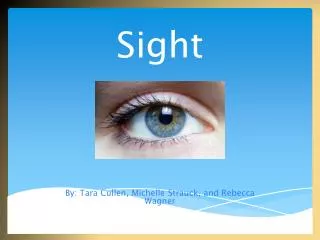
Sight. By: Tara Cullen, Michelle Strauck , and Rebecca Wagner. The Main S tructures of the Eye. Eyelids : acts as a preventive covering for the eyeballs while helping to keep its surface lubricated. It meets at the medial and lateral corners of the eye.

Sight
E N D
Presentation Transcript
Sight By: Tara Cullen, Michelle Strauck, and Rebecca Wagner
The Main Structures of the Eye • Eyelids:acts as a preventive covering for the eyeballs while helping to keep its surface lubricated. It meets at the medial and lateral corners of the eye. • Eyelashes: help trap debris and prevent unwanted materials from entering the eye, and the lid themselves block excess light and foreign objects. • Conjunctiva: is a thin, transparent mucous membrane that covers three parts of the eye. It lines the eyelids and covers part of the outer surface of the eyeball. Generally, the conjunctiva makes blinking and eye movements comfortable. • Sclera: is the tough, white outer layer of the eyeball. It begins at the edge of the cornea and surrounds the eyeball. This is made of tightly woven interlocking fibers which protect the eyeball from injury and help it to hold its spherical shape.
Structures Cont’d. • Cornea: is the clear, round, central window in the front of the eyeball which light travels through to enter the eye. It protects the eye, allows light to enter through the eye, and bends and refracts light so that images can focus on the retina and travel to the brain. • Iris: is the colored, circular part of the eye that forms the pupil in its center. It’s made up of muscles and tissues that adjust the size of the pupil. • Pupil: is defined as the dark appearing round space in the middle of the iris. The darker it is, the larger the pupil will get to let in more light. • Lens: this refracts or bends light so that an image can form on the retina for the brain to see. The lens is made of proteins that form a crystal-like structure, resulting in a clear lens that allows light to pass. • Retina: is a thin layer of complex nerve tissue that lines the inside back wall of the eyeball. It receives images made of light rays, transfers those images into electrical signals, and sends the signals to the brain to be seen. • Optic nerve:carries electrical signals from the retina toward the part of the brain known as the visual cortex. It carries information from the eye to the brain, where that information can be processed into an image that is seen.
How the Eye Works • The first step in how your eye works is the light from the outside world travel your eye. • The light than enters the eye through the pupil and travels to the cornea. • The cornea then focuses the light upon the lens. • The lens further focuses the light on the retina. The image is flipped over and spread across the back of the retina. The retina then carries the light signals to the brain via the optic nerve. • The pathway of light through the eye is cornea aqueous humor (through pupil) aqueous humor lens vitreous humor retina
The Eye and Vision • The external/accessory structures of the eye: • Extrinsic eye muscles aim the eye for following moving objects • The lacrimal apparatus produces a saline solution • Eyelids protect the eyes • Conjunctiva is a mucous membrane covers the anterior eyeball • Three layers that form the eyeball: • The sclera forms most of the outer, tough, protective, fibrous layer • The vascular layer , or middle coat provides nutrition to the internal eye structures • The sensory layer consists of the two-layered retina, which is the pigmented epithelium and the innermost coat which contains photoreceptors
Color Blindness Trouble seeing red, green, or blue or a mix of these colors Makes it harder to learn, read and prevent some from certain careers
Causes of Colorblindness Most cases are genetic; others caused by aging, injury to the eye, or side affects to medication People usually have three color cones that sense red, green, or blue light found in the central part of the retina Color blindness is when you are missing one of these cone cells or one is not working correctly Men are more susceptible to color blindness due to the sex-linked X-chromosomes and they only carry one X chromosome Women carry two X chromosomes so its more difficult to carry it on to them
Symptoms You are still able to see many colors but you may not be aware you aren’t seeing the same as others You are able to see shades of regular colors meanwhile others can see up to thousands of colors In rare cases, some can only see black in white
Vision disorders About a quarter of the population is nearsighted. They can see things up close, but not in the distance. This is called myopia and is caused by an eyeball that is too long or a cornea that is too steep. Many people have difficulty seeing things that are close up. For some this is a life-long problem that is cause by an eyeball that is too short or a cornea that is too flat. The condition is called hyperopia, often referred to as a “farsightedness.”
Disorders Cont’d. Most people will develop problems seeing up close as they age. This is called presbyopia. It is the inability to adjust focus to things close up due to weakening of the eye muscles and loss of flexibility in the eye. Unlike hyperopia, presbyopia does not enhance distance vision. People who are nearsighted remain nearsighted as they lose their close vision with age. People with astigmatism have a football shaped cornea which creates multiple focal points, causing blurred vision. It can be present in combination with myopia, hyperopia, and presbyopia adding to vision problems. In cases where astigmatism only affects near or far sighted vision it is often mistaken for near or far sightedness.
Corrective Laser Eye Surgery http://www.youtube.com/watch?v=f-YkzgfgN2k Also known as LASIK Used to correct vision in people who are nearsighted, farsighted and who have astigmatism LASIK works by reshaping the cornea to make the light traveling through the cornea focused properly
Advantages of Laser Eye Surgery 80% of patients get their eyesight back to normal There is virtually no pain The surgery takes as little as 15 to 20 minutes Vision is corrected almost immediately
Disadvantages of Laser Eye Surgery The surgery can not be reversed Any changes have to be made with more surgery If an error occurs during surgery, vision can be altered completely and permanently
Side Effects Dry Eyes Glares Scarring of the cornea Red patches Fluctuating vision
Diseases Cataracts – clouded lenses Glaucoma – damage to the optic nerve from too much pressure in the eye Retinal disorders – problems with the nerve layer at the back of the eye Conjunctivitis – an infection also known as pink eye