Define the Following Vocabulary
440 likes | 671 Vues
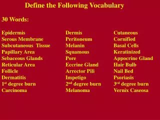
Define the Following Vocabulary. 30 Words: Epidermis Dermis Cutaneous Serous Membrane Peritoneum Cornified Subcutaneous Tissue Melanin Basal Cells Papillary Area Squamous Keratinized Sebaceous Glands Pore Appocrine Gland Reticular Area Eccrine Gland Hair Bulb

Define the Following Vocabulary
E N D
Presentation Transcript
Define the Following Vocabulary 30 Words: Epidermis Dermis Cutaneous Serous Membrane Peritoneum Cornified Subcutaneous Tissue Melanin Basal Cells Papillary Area Squamous Keratinized Sebaceous Glands Pore Appocrine Gland Reticular Area Eccrine Gland Hair Bulb Follicle ArrectorPili Nail Bed Dermatitis Impetigo Psoriasis1st degree burn 2nd degree burn 3rd degree burn Carcinoma Melanoma VernixCaseosa
Integumentary System • One of our 11 organ systems. • Consists of the skin (i.e., the cutaneous membrane) plus all the appendages (or accessory structures) of the skin including: • Sweat glands (sudoriferous glands) • Sebaceous glands (oil glands) • Hair • Nails
General Functions of the Integumentary System • Protection from mechanical injury • Physical protection of pathogen entry • Chemical prevention of pathogen entry • Sensation • Thermoregulation • Metabolic functions • Looking good
Basic Skin Structure • The skin has 2 main layers: • The superficial, avascular epidermis (epi means “above” and dermis means “skin”) consisting of 4-5 layers of epithelial cells resting upon a basement membrane. • The deep, vascular dermis consisting of fibrous connective tissue. Contains multiple blood vessels, and the accessory appendages.
Identify the epidermis and the dermis! Which is made of connective tissue? What type?
The Epidermis • Keratinized stratified squamous epithelium. • Avascular. • Consists of 4 distinct cell types arranged in 4 or 5 distinct layers. • Epidermal cell types: • Keratinocytes (structure) • Melanocytes (pigment) • Merkel cells (sensation) • Langerhans’ cells (phagocytes - immune defense) Yellow arrow indicates the epidermis of thick skin
Keratinocytes • Most numerous epidermal cell – found in all layers of the epidermis. • Chief function is the production of keratin – a tough fibrous protein that gives strength and confers a lot of protective ability. • Tightly connected to one another by desmosomes. • Provides continuity, strength, and protection. • Is the reason skin flakes off in sheets rather than as individual cells. Almost all of the epidermal cells in this slide are keratinocytes
Keratinocytes • New cells are continuously made in the deepest layer pushing the older cells up. • As the keratinocytes move farther from the deepest layer, they make the keratin that eventually dominates their cell contents. When they have reached the upper layer, they are nothing more than scale-like bags of keratin.
Melanocytes • Spider-shaped epithelial cells that synthesize the protein pigment melanin. • Found in the deepest layer of the epidermis. • Melanin is made and then packaged into membrane-bound granules called melanosomes. • Granules are transferred to the keratinocytes in the 2 deepest layers of the epidermis. Arrows indicate 2 melanocytes.
Melanocytes • Melanin granules accumulate on the “sunny side” of the nucleus of the keratinocytes. • Would that be the apical or the basal side? • Melanin granules protect the DNA within the nucleus from being damaged by the ultraviolet radiation from the sun.
Skin Types • Thick Skin • Found on soles of feet and palms of hands and corresponding parts of fingers and toes. • Contains 5 epidermal layers or strata (“sheets”): • Stratum basale • Stratum spinosum • Stratum granulosum • Stratum lucidum • Stratum corneum
Skin Types • Thin skin • Found everywhere else on the body. • Contains only 4 layers. (lacks a stratum lucidum). • The remaining 4 layers are thinner than those of thick skin. • Why is thick skin found on the palms and soles? What is the advantage of that? Note: this slide is at a higher mag. than the thick skin slide on the previous page
Notice the 4 layers of thin skin in both the cartoon and the photomicrograph.
Stratum Basale • Single row of cuboidal keratinocytes with melanocytes Merkel cells interspersed. • Cells in this layer are highly mitotic – they’re dividing often. Due to this fact, this layer is a.k.a. the stratum germinativum. • Deepest epidermal layer. Firmly attached to the underlying dermis.
Stratum Spinosum • 2nd deepest layer. Consists of 8-10 layers of cells. • Cells of the lower layers can still be mitotic. • As cells get pushed upward, they begin to flatten and begin to make the precursors of keratin. • A.k.a. the “prickly layer,” because in tissue sections, they shrink and pull back. This makes their exposed desmosomes connecting adjacent cells appear to resemble spikes or spines. Do you see the spines?
Stratum Granulosum • 3-5 cell layers thick. • Cell morphology begins to change dramatically as cells continue to flatten and their nuclei and organelles disintegrate. • Accumulate granules containing a precursor of keratin and granules containing a waterproofing agent. • If water can’t diffuse upward, how would the cells above this layer receive nutrients? What would happen to them?
Stratum Lucidum • 3-5 layers of flat, dead keratinocytes. • Appears clear in the light microscope because it lacks nuclei and organelles which typically stain well.
Stratum Corneum • Outermost stratum. 20 – 30 layers of flat (squamous), highly keratinized, dead cells. • Protects against mechanical abrasion – cells can absorb impacts and simply flake off if necessary. Prevents pathogen entry. Prevents desiccation (drying out). • A.k.a. the cornified layer. • The process by which cells in the stratum basale divide and then advance upward becoming more and more keratinized and less and less alive as they go is known as cornification.
Skin Color • Due to 3 pigments: • Melanin • Carotene • Hemoglobin • Of these, only melanin is made in the skin. • Melanin: • Polymer of tyrosine amino acids. Its synthesis is catalyzed by an enzyme called tyrosinase. Albinos lack this enzyme. • Ranges in color from yellow to reddish brown to black. • All people have the same # of melanocytes, individual variations in skin color are due to how much and what type of melanin is made. • Freckles and moles are local accumulations of melanin.
Carotene • Yellow to orange pigment found in plant products such as carrots. • When large amounts are eaten, it can be deposited in the stratum corneum of thick skin. • Hemoglobin • Pigmented protein that transports oxygen within the blood. • In Caucasians, the fair skin allows the crimson color of oxygenated blood to make the skin have a somewhat pinkish hue.
Dermis • Strong, flexible fibrous connective tissue. • Divided into papillary dermis and reticular dermis. • Papillary dermis is the upper 1/5 of the dermis and consists of loose (areolar) CT. • Provides an arena for immune cells to fight invaders. • Projects upward (as dermal papillae) to interdigitate and form a strong connection with the epidermis. • Heavily invested with blood vessels – they constrict in cold weather and dilate in warm weather. Why? • Also contains multiple sensory receptors.
Dermis • Reticular dermis is lower 4/5 and consists of dense irregular connective tissue. • The prominent, horizontally running collagenous fibers give the skin strength and resiliency. • Elastin gives the skin the ability to stretch and recoil. • The majority of the appendages of the skin are contained within the dermis. Red arrow indicates the papillary dermis and blue arrow indicates the reticular dermis
Appendages of the Skin • Sebaceous glands • Sudoriferous glands • Ceruminous glands • Hair • Nails 1 4 2
Sebaceous Glands • Simple alveolar glands found everywhere except palms of the hands and soles of the feet. • Do the ducts of these glands branch? • Are these glands exo- or endocrine? • Secrete an oily, lipid-rich secretion called sebum. • Lanolin is actually sheep sebum • Sebum is typically secreted into a hair follicle or occasionally onto the body surface. • Sebum softens and lubricates the skin. It also decreases the skin’s permeability to water and is quite bactericidal.
The sebaceous gland is indicated by the arrow. Note how its duct is unbranched and how it empties into a hair follicle.
Sudoriferous Glands • Sweat glands. • Distributed over the entire body except the nipples and portions of the external genitalia. • Over 2.5 million per person. • 2 types: • Merocrine sweat glands • Apocrine sweat glands
Merocrine Sweat Glands • More numerous than apocrine sweat glands. • Especially prominent on the palms, soles, and forehead. • Simple, coiled, tubular glands. • Duct empties into a funnel-shaped pore at the skin surface. • Major function of merocrine sweating is to cool the body – thermoregulation.
Merocrine Sweat Glands • Merocrine sweat is a dilute watery solution of some salts (including NaCl), vitamin C, antibodies, small amounts of nitrogenous wastes (urea, uric acid, and ammonia), and lactic acid. • pH of sweat is 4-6 creating a film on the body known as the acid mantle. Such an acidic environment is bacteriostatic – prevents bacterial reproduction and growth.
Apocrine Sweat Glands • Found primarily in the axillary, pubic, and anal regions of the body. • Also found in the facial region in men only. • Larger then merocrine sweat glands. • Ducts empty into hair follicles. • Apocrine sweat is thicker than merocrine sweat since it contains more lipids and proteins. • When it’s degraded by epidermal bacteria, foul odors can be produced.
Apocrine Sweat Glands • Become active at puberty. • Secrete sweat during times of pain, stress, and sexual activity. • May (??) have a possible pheromone-like function. • Play no role in thermoregulation. Remember: Glands are derivatives of epithelial tissue. So even though many reside in the dermis, they are not made from connective tissue.
Ceruminous Glands • Modified apocrine glands found in the inner lining of the external ear canal. • Secrete a sticky waxy substance called cerumen (earwax). It combines with sebum and dead epidermal cells to form a bitter compound that offers a barrier to entry of the ear.
Hair and Hair Follicles • Hair is a flexible strand made of highly keratinized dead cells. • The keratin in both hair and nails is hard keratin, a stiffer, more compact version of the soft keratinthat dominates the epidermis. It is tougher and its cells do not individually flake off. • The hair is made by the living hair follicle.
Hair and Hair Follicles • Hair consists of a shaft which protrudes from the skin and the root which is within the skin. At the base, the root gets wider forming the hair bulb. • The hair consists of 3 concentric regions: • Medulla the innermost region consisting of large cells and air spaces. • Cortex surrounds the medulla and consists of flattened keratinized cells • Cuticle outer layer of flat keratinized cells that overlap like shingles with their free edges pointing upward. What word is misspelled in this picture?
Hair and Hair Follicles • The hair follicle surrounds much of the hair root. • It contains an outer connective tissue sheath and an inner epithelial root sheath. • At the base of the hair follicle is a single layer of mitotic cells derived from the stratum basale. This is the hair matrix. • All the cells of the hair are derived from the hair matrix. Just beneath the hair matrix is an obvious dermal papilla called the hair papilla. It contains the blood vessels that nourish the matrix and the cells of the hair follicle.
Notice the hair shaft, hair follicle, papilla, and the multiple sebaceous glands.
Hair and Hair Follicles • Wrapped around the bulb of the follicle is a network of sensory nerve endings known as the hair root plexus. Allow the hairs to serve a sensory function. • Attached to each hair is a bundle of smooth muscle known as an arrector pili muscle. In times of fright or cold, these muscles contract and cause the hair to stand on end – and produces goose bumps. • Increases airflow in mammals with significant hair (i.e., not humans) and increases the apparent size of an animal with significant hair. Vestigial in humans.
The arrow indicates an arrector pili muscle. In this picture, you should also try to identify the shaft, root, follicle, hair papilla, and sebaceous gland.
Subcutaneous Tissue • A.k.a. the hypodermis or the superficial fascia. • Deep to the dermis. • Consists of primarily adipose tissue plus some areolar connective tissue. • Stores energy, provides insulation and padding. • Anchors the skin to underlying structures, especially muscles. • Different distribution between the sexes.
Skin Cancer • Because of its role as our external covering, the skin takes a tremendous amount of abuse. • One serious disorder that can result is skin cancer. • Cancer can be thought of as uncontrolled cell division and growth. • There are 3 types of skin cancers we will discuss: • Basal cell carcinoma • Squamous cell carcinoma • Malignant melanoma From abnormal cells, a cancerous cell develops Cancerous cells spread, forming a tumor An abnormal cell develops
Skin Cancer • Basal cell carcinoma • Most common (70% of skin cancers) • Least vicious • Usually cured via surgical removal • Consists of uncontrolled growth of cells of the stratum basale. They’ll proliferate and invade the dermis and hypodermis. • Often occurs on sun-exposed areas of face and neck
Skin Cancer • Squamous cell carcinoma • Arises from keratinocytes of stratum spinosum. • 25% of cases. • Good prognosis if caught and treated early (surgical excision or radiation). • Can be fatal if it metastasizes to the lymph nodes.
Skin Cancer • Malignant melanoma • Least common and most dangerous. • Cancer of melanocytes. • Often arises from a pre-existing mole. • Follow the ABCD rule for early detection: Asymmetry (2 sides do not match) Border irregularity Color (multiple) Diameter (>6mm is bad!)