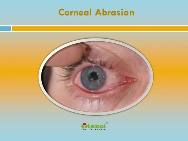
6 layers of cornea Response of cornea to injury Abrasion vs ulcer Corneal opacity- grades
Re-cap of last class. 6 layers of cornea Response of cornea to injury Abrasion vs ulcer Corneal opacity- grades Corneal transparency- causes. Keratitis - Bacterial. Dr. Soujanya K MBBS, MS, DNB, FPRS Assistant Professor YMCH. Specific learning objectives.

6 layers of cornea Response of cornea to injury Abrasion vs ulcer Corneal opacity- grades
E N D
Presentation Transcript
Re-cap of last class • 6 layers of cornea • Response of cornea to injury • Abrasion vs ulcer • Corneal opacity- grades • Corneal transparency- causes
Keratitis- Bacterial Dr. Soujanya K MBBS, MS, DNB, FPRS Assistant Professor YMCH
Specific learning objectives • Classification of keratitis • Predisposing factors for bacterial keratitis • Clinical features • Stages of bacterial keratitis • Complications ans sequel of bacterial keratitis • Management of bacterial keratitis
Keratitis Classification: • Based on location: • Superficial • Deep
Keratitis Classification: • Based on Aetiology: • Infectious • Non- infectious • immune-mediated • degenerative • neoplastic • traumatic (including chemical and thermal injuries).
Normal cornea Highly virulent organisms usually NOT present Tear film & resistance of normal tissue protects against infection
Bacteria causing keratitis • Organisms which invade de-epithelised or injured cornea- • Staphylococci • Pneumococci • Enterobacteriaceae • Atypical mycobacteria
Bacteria causing keratitis • Organisms which invade intact cornea- • Gonococci • Diphtheria bacilli
Y DRY EYE CONTACT LENS USE TRAUMA KERATOMALACIA
Photophobia Lacrimation Diminution of vision
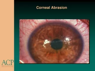
Circum corneal congestion Corneal Infiltration + saucer shaped ulcer Hypopyon
Suppurativekeratitis • Due to organisms that produce toxins which cause tissue death (necrosis) and pus formation in the corneal tissue. • Nearly always exogenous
localized necrosis of the anterior layers of the cornea Epithelium desquamates & Bowman’s membrane -damaged
Desquamated tissue adheres to ulcer floor
Progressive stage Walls: project above the normal surface of the cornea owing to swelling caused by the fluid imbibed by the corneal lamellae. Surrounding area is packed with leucocytes and appears as a greyzone of infiltration.
Some of the toxins produced by the bacteria diffuse through the cornea into the anterior chamber Exert an irritative effect upon the vessels of the iris and ciliary body resulting in keratouveitis. If the irritation is great, leucocytosis takes place, and polymorphonuclear cells poured out by the vessels pass into the aqueous and gravitate to the bottom of the AC & form hypopyon
Regressive stage Line of demarcation forms : wall of polymorphonuclear leucocytes
Surrounding infiltration & swelling disappear, floor and edges become more smooth and transparent
vascularization • Restore the loss of substance • Supply antibodies • Play an important role in resolving bacterial infections
The healing of a corneal ulcer Cicatrization: Regeneration of collagen and the formation of fibrous tissue. The newly formed fibres are not arranged regularly, hence they refract the light irregularly and the scar is therefore opaque
Progressive stage Regressive stage Vascularization and scaring
Quick revision • Classification of keratitis • Predisposing factors for bacterial keratitis • Clinical features • Stages of bacterial keratitis
Ectatic cicatrix Marked thinning of the entire cornea at the site of the ulcer so that it bulges under the influence of the normal intraocular pressure secondary keratectasia
Some ulcers extend rapidly in depth uptoDescemet's membrane