Sense Organ
Sense Organ. Jun Zhou ( 周俊 ), M.D. & Ph.D. School of Medicine, Zhejiang University . 20121218. LEARNING METHODS Listen attentively and think actively during the lecture. Preview and review the textbook and atlas as much as you can.

Sense Organ
E N D
Presentation Transcript
Sense Organ Jun Zhou (周俊), M.D. & Ph.D. School of Medicine, Zhejiang University 20121218
LEARNING METHODS • Listen attentively and think actively during the lecture. • Preview and review the textbook and atlas as much as you can. • NEVER passing by a word without knowing its definition. • To understand the structure and function of each organ, not just memorize them. • Email: zhjjwm300@zju.edu.cn
Special sense receptors • Responsible for the five special senses: taste, smell, seeing, hearing, feeling • Tranduce stimuli from the environment into electrical impulses
Specialized diffuse receptors • Two important sensory organ • The eye • The ear
Specialized diffuse receptors • Free nerve terminals -- feel cold, hot, pain and slight touch • Encapsulated nerve ending --have CT capsule • Pacinian corpuscle • Meissner corpuscle • Proprioceptive receptors
Meissner corpuscle • Ellipsoid, encapsulated receptor • Located in the dermal papillae of thick skin • Fine touch perception
Pacinian corpuscle • Large ellipsoid encapsulated receptor • Located in the dermis. hypodermis, mesenteries • Multilayer capsule surrounds inner unmyelinated nerve terminal • Perceive pressure, vibration
Muscular spindles Proprioceptive receptors • 3-12 small encapsulated intrafusal muscle fibers • Sense differences in muscle length and tension
Two special sensory organs The eye is a complex sensory organ that provides the sense of sight.In many ways, the eye is similar to a digital camera.It capture and focus light, convert the light image into neuronal impulse, then transmit to the brain via the optic nerve. The ear is a three-chambered sensory organ that functions as an auditory system for sound perception and as a vestibular system for balance. • Eyes: visual organ • Ears: the organ of hearing and equilibrium.
Fibrous layer Walls Vascular layer eyeball Retina eye Content:Aqueous humor、Lens、 Vitreous body Accesory structure:Eyelid、Muscles of the eye、Lacrimal gland
Eyeball Walls Fibrous layer, includes the cornea, covers the anterior one sixth of the eye,the transparent portion. It has a prominence or convexity. The sclera is composed of dense fibrous connective tissue that provides attachment for the extrinsic muscles of the eye.The sclera has a slightly blue in children because of its thinness and is yellow in the eldly because of the accumulation of lipofuscin. Blood vessels and melanin pigment give the choriod an intense dark-brown color.Provides nutrients to the retina.The anterior forms the ciliary body and iris.The ciliary body is a ringlike thickening that extends inward.The iris is a contractile diaphragm that extends over the anterior surface of the lens. The central circular aperture is the pupil. The retina consists largely of photoreceptors cells,forming visual impulses along optic nerve. Cornea1/6 Fibrous layers Eye ball Vascular layers Retina Sclera 5/6 Choroid2/3 Ciliary body Iris
Cornea The transparent cornea is only 0.5mm thick at its center and about 1mm thick peripherally.It consists of three cellular layers ,which are separated by two homogeneous membrane. Colorless, transparent 5 layers: epithelium Bowman’s membrane Stroma Descemet’s membrane endothelium
Cornea a.epithelium: • Non-keratinized stratified squamous epi. • 5-6 layers • Numerous mitotic figures • No vessels. • Free nerve ending b.Bowman’s membrane( anterior basement membrane) • An acellular homogeneous membrane (collagen fibrils) • Stability & strength, no regeneration
C. Stroma or substantia propria • Several lamellae of fine collagen f.network • Flattened fibroblasts • G.S.rich in chrodroitin sulfate D. Descemet’s membrane (posterior limiting lamina) • Acellular homogeneous membrane • Can be repaired by endothelial cells E.Endothelium • Like mesothelium in its morphology • Regulate the water content of the stroma maintain transparency
The reasons of cornea transparent • No blood vessels & pigments • Basal of epi. is plane • Uniform spacing of collagen fibrils and lamellae in stroma • G.S. with transparent nature & maintains proper water
Retina • In the neural retina, two regions or portions that differ in function are recognized: • The nonphotosensitive region (nonvisual part),located anterior to the ora serrata, lines the inner aspect of the ciliary body and the posterior surface of the iris. • 2) The photosensitive region (optic part) lines the inner surface of the eye posterior to the ora serrata except where it is pierced by the optic nerve. Two regions: • The nonphotosensitive region (nonvisual part) Located anterior to the ora serrata, no photoreceptors. • The photosensitive region (optic part) Lines the inner surface of the eye posterior to the ora serrata (except the optic papilla)
Retina The retina, is the innermost of the three concentric layers of the eye.It consists of two basic layers: 1)RPE, the outer layer that rests on and is firmly attached through the Bruch’s membrane to the choriocapillary layer of the choriod 2)Neural retina or retina proper, the inner layer that contains the photoreceptors A potential space exists between the two layers of the retina. The two layers may be separated mechanically in the preparation of histologic specimens. Separation of the layers, “retinal detachment”, also occurs in the living state as a result of eye disease or trauma. 4 layers of cells: • Pigment cells • Optic cells • Bipolar cells • Ganglion cells
Junctional complex consisting of gap junctions and elaborate zonulae occludentes and adherentes. This junctional complex is the site of the blood-retina barrier. • 2) Processes project for a short distance between the photoreceptors of the rods and cones. Numerous elongated melanin granules are present in many of these processes.They aggregate on the side of the cell nearest the rods and cones and are the most prominent feature of the cells. • The RPE serves several important functions including: • Absorption of light passing through the neural retina to prevent reflection • 2) Isolation the retinal cells from blood-borne substances.It serves as a major component of the blood-retina barrier via tight junctions between RPE cells. • 3) Phagocytosis and disposal of membranous discs from the rods and cones of the retinal photoreceptor cells. • 4)Participation in restoration of photosensitivity to visual pigments that dissociated in response to light. The metabolic apparatus for visual pigment resynthesis is present in the RPE cells. Pigment epithelium • Structure: • 1) Simple cuboidal epi. Attached to choroid and easy separated from retina (detachment of retina) • 2) Junctional complex , • 3) Melanin granules • 4) Processes (contain pigment granules) • Function: • absorb light,protect rod and cone from strong light • Blood-retina barrier • 3) Phagocytize the membranous discs from retinal photoreceptor cells • 4) Store vitamin A to assist in forming rhodopsin
Optical cells The rod and cones are the outer segment of photoreceptor cells .The light that reaches the photoreceptors must first pass through all of the internal layers of the neural retina.The rods and cones are arranged in a palisade manner; therefore, in the light microscope, they appear as vertical striation. • bipolar neurons • The rods and cones • Glial cells(Müller cell)
Rod cell Outer segment of the photoreceptor is roughly cylindrical,(hence, the descriptive name rod). With the TEM, 600 to 1000 regularly spaced horizontal discs are seen in the outer segment. The membranous discs are formed repetitive transverse infolding of the plasma membrane in the region of the outer segment near the cilium.Rods form new discs throughout their life span. Discs are formed in cones in a similar manner but are not replaced on a regular basis. Rod discs lose their continuity with the plasma membrane from which they are derived soon after they are formed, eventually shed and phagocytosed by the pigment epithelial cells. Rhodopsin in rod cells initiates the visulal stimulus when it is bleached by light. It is a derivative of vitamin A. Thus, an adequate intake of vitamin A is essential for normal vision. Prolonged dietary deficiency of vitamin A leads to the inability to see in dim light (night blindness) • Thin,elongated cells, about 120 million rods • A body and two opposite processes • Outer segment and inner segment • separated discs ,shed disc phagocytized by pigment cells • rhodopsin (visual purple) Function: • sensitive to low intensity light • Night vision (lack of vitamin A leads night blindness)
cone cell Outer segment of the photoreceptor is roughly conical.(hence, the descriptive name cone). Discs are formed in cones in a similar manner but are not replaced on a regular basis. Discs within the cones retain their continuity with the plasma membrane. The specificity of the cones provides a functional basis to explain color blindness.True color-blind individuals (almost all are male) are dichromats and are believed to have a defect in either the red-, green-, or (much less commonly) blue-sensitive cones. • About 7 million cells • Located in posterior part of retina,especially in fovea • Outer and inner segments (conical) • Continuous discs & not renewed Function • 1)sensitive to high intensity light • 2)color distinguishing(red、blue、green iodopsin)
Bipolar cells Interconnected cells is reduced to provide greater visual acuity. Four types of conducting cells---bipolar,horizontal, interpexiform and amacrine cells. • An axon & a dendrite • Synapse with photoreceptor cells and ganglion cells Müller cells • Extend entire thickness of retina • Neuroglia Horizontal cells Amacrine cells Interconnected cells—provideing greater visual acuity
Ganglion cells These nerve cells have lightly staining round nuclei with prominent nucleoli and have Nissl bodies in their cytoplasm. Axonal process passes into the nerve fiber layer, then goes into the optic nerve. • The dendrite synapse with bipolar cells • The axons concentrate together form optic nerve
Macula lutea is the area surrounding the fovea, is yellowish because of the presence of yellow pigment.Here, the retinal cells and their processes, especially the ganglion cells, are heaped up on the side of the fovea so that light may pass unimpeded to this most sensitive area of the retina. Specilized regions of the retina • Ora serrata :neural layer ends anteriorly at ciliary body,pigment cells extend to cover posterior iris • Macula lutea:directly on eye’s posterior pole. “yellow spot”,mostly cones • Fovea centralis: central pit of macula,only cones, vision acuity straight on • Optic disc:blind spot,no rods or cones,optic nerve exits,medial and inferior to fovea centralis.
10 layers 1 = pigmented epithelium 2 = layer of photoreceptors 3 = external limiting membrane 4 = outer nuclear layer 5 = outer plexiform layer, where photoreceptors synapse 6 = inner nuclear layer of bipolar neurons 7 = inner plexiform layer, where bipolar neurons synapse with ganglion cells 8 = ganglion cell layer 9 = optic nerve layer 10 = internal limiting membrane The specific arrangement and association of the nuclei and processes of these cells result in the retina being organized in ten layers that are seen with the light microscope. The ten layers of the retina, from outside inward, are 1.RPE: the outer layer of the retina 2.contains the outer and inner segments of photoreceptor cells 3.the apical boundary of muller’s cells 4.contains the cell bodies(nuclei) of retinal rods and cones 5.contains the processes of retinal rods and cones and processes of the horizontal.amacrine and bipolar cells that connect to them. 6.contains the cell bodies of horizontal, amacrine, bipolar and muller’s cells. 9.processes of ganglion cells that lead from the retina to the brain 10.composed of the basal lamina of Muller’s cell.
Visual pathways light cornea champer lens vitreous bodyretina pigment epithelium rods and cones bipolar cells ganglion cells optic nerve fibers
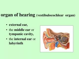
The ear is a three –chambered sensory organ, functions as an auditory system for sound perception and as a vestibular system for balance. There are three divisions of ear. The external and middle ear collect and conduct sound energy to the internal ear, where auditory sensory receptors convert that energy into electrical impulses. The sensory receptors of vestibular system are located in the internal ear.These receptors respond to gravity and movement of the head. Ear • External, middle, inner ear • External and middle ear: • gathers and funnels sound waves • Inner ear: • sensory of hearing and balance
Mastoid process (auricle) External: 1)auricle(pinna):oval,projects from the lateral surface of the head.Its shape is determined by internal supporting elastic cartilage. 2)an air-filled tubular space that follows a slightly S-shaped course 3)The tympanic membrane separates the external acoustic meatus from the middle ear. Middle: 1)an air-filled space, called typanic cavity, which are spanned by three small bones. 2)The auditory tube connects the middle ear to the nasopharynx. Equalizing the pressure of the middle ear with atmospheric pressure.It is common for infections to spread from pharynx to the middle ear via the auditory tube. 3)Mastoid bone is spongy bone. • External ear : • auricle • external acoustic meatus • tympanic membrane (eardrum) • Middle ear: • tympanic cavity • auditory tube • mastoid process
Internal ear Bony labyrinth Membranous labyrinth The internal ear consists of two labyrinthine compartments, one contained within the other. The bony labyrinth is a complex system of interconnected cavities and canals in the petrous part of the temporal bone. The vestibule is small oval chamber located in the center of the bony labyrinth.The semicircular canals extend from the vestibule posteriorly, and the cochlea extends from the vestibule anteriorly. The semicircular canals are tubes within the temporal bone that lie at right angles to each other. The cochlea is a cone-shaped helix connected to the vestibule. The membraneous labyrinth consists of a series of communicating sacs and ducts containing endolymph. It is suspended within the bony labyrinth, and remaining space is filled with perilymph. Endolymph has similar composition to intracellular fluid.Perilymph similar to extracellular fluid. • Bony labyrinth: • Semicircular canals • Vestibule • Cochlea • Menbranous labyrinth: • Cochlear labyrinth • Vestibular labyrinth • three semicircular ducts • Utricle and saccule
Specialized sensory cells are located in six regions in the membranous labyrinth. 1)cristae ampullaris are located in the membranous ampullae of the semicircular ducts.They are sensitive to angular acceleration of the head. 2)Two maculae,sense the position of the head and its linear movement. 3)functions as the sound receptor. • Six sensory regions of the membranous labyrinth: • Three crista ampullaris • Two maculae(maculae of utricle, maculae of saccule) • The spiral organ of Corti
Crita ampullaris Project from the wall of the membranous labyrinth into the endolymphatic space. 1)cupula: a gelatinous protein-polysaccharide mass, attached to the hair cells of each crista. 2)The stereocilia and kinocilium of each hair are embedded in the cupula. Rotational movement move the semicircular canal and membranous semicircular ducts.Deflection of the stereocilia of the hair cells generates nerve impulses in the associated nerve ending. three,located in the membranous ampullae of the semicircular ducts Composition: Supporting cells:support,forming cupula Sensory hair cells:with stereocilia and kinocilium are embedded in the cupula Function: sensors of angular acceleration of the head
The maculae of the utricle and saccule are oriented at right angles to one another.One is in a horizontal plane, another one is in a vertical plane. • Maculae of utricle and saccule: • Located in the vestibule • Sense the position of the head and its linear movement
The cochlea duct divides the cochlear canal into three parallel compartment or scalae.The cochlea duct itself is the scala media. The scala vestibuli and scala tympani are the spaces above and below, respectively. • Note: • The position of cochlear duct within the bony cochlea • The scala vestibuli and the scala tympani, containing perilymph • The scala media containing endolymph
Schematic diagram of the cochlea: The scala media is the triangle space with its acute angle attached to the modiolus.Both the scala vestibuli and the scala tympani are filled with perilymph, whereas the cochlear cduct is filled with endolymph.The spiral organ of Corti rests on the lower wall of the scala media. The scala vestibuli and the scala tympani are perilymph-containing spaces that communicate with each other at the apex of the cochlea through a small channel. Scala vestibuli Scala tympani
Corti’s organ:sensor of sound vibration The tectorial membrane extends from the spiral limbus over the cells of the spiral organ of Corti.And attaches to the stereocilia of the hair cells. Between the inner pillar cells and outer pillar cells, form a triangular tunnel. On the lower wall of the scala media Tectorial membrane Inner (close to spiral lamina)and outer (farther from the spiral lamina) hair cells Supporting cells: inner and outer phalangeal cells pillar cells
Sounds Pathawy • Sound comes • Hits tympanic membrane to vibrate • three auditory ossicles vibrate • vibration at tympanic (oval) window • Vibration in the perilymph of the scala vestibular to the scala media • Vibrates of basilar membrane and tectorial membrane,and hair cells attached to also vibrates • Vibrate the stereocilia of the hair cells and initiate neuronal transduction High-frequency sounds cause maximal vibration of the basilar membrane near the base of the cochlea;low-frequency sounds cause maximal displacement nearer the apex.
Clinical Correlation • Vertigo: dysfunction of vestibular system Causes: viral infections, certain drugs, tumors, excessive stimulation (seasickness, carsickness, or airsickness) • Hearing loss 1)Conductive hearing loss:sound waves are mechanically impeded from reaching the auditory sensory receptors within the internal ear.such as excessive accumulation of cerumen. 2)Sensorineural hearing impairment:injury to the auditory hair cells or the cochlea nerve. May be congenital or acquired. Causes include infections, trauma (exposure to excessive noise), administration of certain antibiotics, aging.
cornea Ciliary body iris lens sclera
OBJECTIVES • Know the general layers of the eye. • Describe the structure of Cornea and its reason of transparent. • Describe the structure of Retina and the function of pigment cell, rod cell and cone cell. • Know the definition of Ora serrata, Macula lutea, Fovea centralis and Optic disc. • Know the general structure of ear. • Describe six sensory regions of the membranous labyrinth and their function.
Sclera Choroid Retina
Scala vestibuli Scala media Scala tympani
Choice: Select the single most appropriate answer. • 1.The posterior wall of eyeball from outside inward contains • A.fibrous layer,vascular layer and retina • B.retina,choriod and sclera • C.choriod,sclera and retina • D.cornea,iris and retina • E.Retina, vascular layer and fibrous layer • 2. The Müller cells of retina belong to • A.sensory neurons • B.neuroglial cells • C.interneurons • D.photoreceptors • E.motor neurons • 3.The cells used color perception and fine visual acuity are • A. ganglion cells B. Müller cells • C. bipolar cells D.rods • E. cones ( A) ( B ) ( E )
( A ) 4.The optic nerve fibers are constituted by axons of A.ganglion cells B.Müller cells C.Bipolar cells D.rods E.Cones 5.The receptor of hearing is located on A.Vestibular membrane B.Crista ampullaris C.Maculae saccule D.Maculae utricle E.Organ of Corti ( E )