Excretion
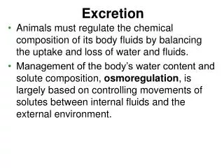
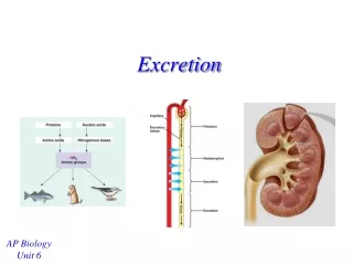
Excretion. Chapter 9. Functions of Excretory Organs . Maintain solute concentrations Maintain body fluid volume Remove metabolic end products Remove foreign substances . Nitrogen Excretion. Nitrogen-based waste compounds derived from proteins and nucleic acids

Excretion
E N D
Presentation Transcript
Excretion Chapter 9
Functions of Excretory Organs • Maintain solute concentrations • Maintain body fluid volume • Remove metabolic end products • Remove foreign substances
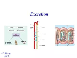
Nitrogen Excretion • Nitrogen-based waste compounds derived from proteins and nucleic acids • Excretion classified by major waste produced • Ammonotelic – ammonia is the principle waste • Ureotelic – urea is the principle waste • Uricotelic – uric acids and urate salts are the principle wastes Fig 27.23
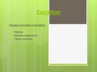
Excretory Processes • Ultrafiltration • Movement of fluid (e.g. blood) through a semipermeable membrane • Membrane allows small particles to pass with the water, large particles (proteins etc.) remain • Active Transport • Movement of solutes against their electrochemical gradients (requires energy) • Secretion – movement of solute into the lumen of the excretory organ • Reabsorption – movement of solute out of lumen
Generalized Excretory Organs • Sponges, Coelentrates and Echinoderms – none • Playhelminths, Nematodes, Annelids – nephridial organs • Crustaceans – antenna glands • Insects – Malpighian tubules • Mollusks and Vertebrates – kidneys
Nephridial Organs • Common in invertebrates • System of tubes • connected to the outside through nephridial pore • Protonephridia • Found in acoelous and pseudocoelous animals (platyhelminths, nematodes, etc.) • Blind-ended tubes with flame cells or solenocytes at closed end • Create current • Draw fluid in from surrounding tissues (filtration) • Water then reabsorbed
Nephridial Organs • Metanephridia • Coelous Animals (e.g., annelids) • Inner cells open into coelomic cavity • Four components: • Nephrostome – funnel-shaped filter • Coiled tubule – secretion and absorption • Bladder – storage • Nephridial pore
Antennal Gland • Crustaceans • Paired glands located in the head • Consist of initial sac, long coiled excretory tubule and terminal bladder • Excretory pore at base of antenna Fig 27.19
Malpighian Tubules • Arachnids and insects • Specialized region of digestive tract • Located btw midgut and hindgut • Blind-ended tubules • Blind ends locates in hemocoel • Some end near rectum Fig 27.21
Malpighian Tubules and Rectum • NO ultrafiltration • Active secretion of K+into lumen • H2O follows passively along osmotic gradient • Content altered by secretion and absorption • Fluid secreted into hindgut • Water and solutes reabsorbed in rectum • Uric acid precipitates Fig 27.22
Molluscan Kidneys • Associated with pericardial cavities • Ultrafiltration from heart • Secretion/reabsorption by renopericardial canal • Stored in bladder (renal sac) • Released into mantle and expelled Fig. 27.20
Vertebrate Kidneys • Ultrafiltration followed by reabsorption • Blood plasma is filtered, then important solutes and water reabsorbed into the blood • 99% of filtered material is reabsorbed • Allows animals to filter out new substances without developing new specialized secretory mechanisms Fig. 27.1
Vertebrate Kidneys • Consists of numerous tubular units called nephrons Figs 27.6
Kidney • Blood delivered into the glomerulus • Tuft of fenestrated capillaries • Site of filtration (blood pressure forces filtered plasma out) • Filtrate collected by Bowman’s capsule Fig 27.1
Kidney • Enters tubular structures • Proximal tubule • Reabsorption of solutes and water • Distal tubule • Further reabsorption and secretion • Collecting duct • Join several distal tubules Fig 27.3
Kidney • Loop of Henle (mammals and birds) • Thin, single loop between proximal and distal tubules • Allows formation of hyperosmotic urine Figs 27.6 and 27.18
Glomerular Filtration • Occurs through fenestrated capillaries • Plasma with small particles filters out • Blood cells and plasma proteins remain • Blood pressure must exceed colloid osmotic pressure Fig 27.1c
Tubular Secretion • Removal of excess ions (K+, Ca2+, Mg2+, H+) • Removal of foreign substances • Active Transport Fig 27.16
Tubular Reabsorption • Active transport of inorganic ions Na+ • Coupled transport of glucose, amino acids, etc. • Osmotic uptake of water Fig 27.16
Hyperosmotic Urine • Mammal kidneys can excrete a hyperosmotic urine • concentrating mechanism occurs in the Loop of Henle • Countercurrent Multiplication • generates osmotic gradient that draws H2O out of the tubules to be reabsorbed • due to active reabsorption of Na+ and Cl-
Loop of Henle • Mechanism • descending limb • permeable to water • ascending limb • impermeable to water • lined w/ ion pumps (Na+ or Cl-) Fig 27.11
Loop of Henle:Ascending Limb • Na and Cl actively transported out of lumen • urea flows out of lumen in thin segment of ascending limb • Creates osmotic gradient Fig 27.14
Loop of Henle:Descending limb • osmotic gradient generated btw interstitial fluid and lumen • H2O moves out of the lumen • Filtrate concentrated to hyperosmotic levels • Water leaving lumen diffuses into the vasa recta (re-enters blood) Fig 27.14
Loop of Henle:Ascending limb • osmotic concentration ’s as solutes are moved out of the filtrate by active Na+ transport Fig 27.14
Collecting Duct • Water flows out as tubule descends into medulla • Water leaving lumen diffuses into the vasa recta (re-enters blood) • Final urine produced is hyperosmotic Fig 27.14
Loop Length and Aridity • Relative length of the loops is longer in animals adapted to dry habitats than in those from wetter habitats Figs 27.8-27.10