Download
1 / 19
220 likes | 512 Vues
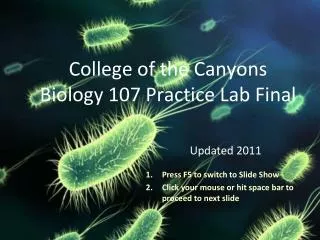
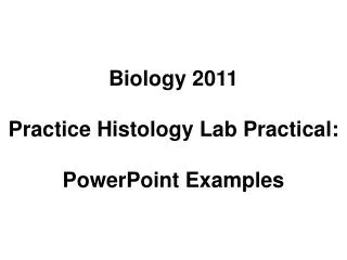
Biology 2021 Practice Final Lab Practical. Each of the following slide images contains a multiple choice question. Bubble in your answers in the appropriate blank circles on your answer sheet.

E N D
Biology 2021 Practice Final Lab Practical Each of the following slide images contains a multiple choice question. Bubble in your answers in the appropriate blank circles on your answer sheet. When asked to “identify an object at the pointer,” this always refers to the object at the very tip of the pointer. No talking or communication of any kind is permitted during the practical. You may raise your hand to consult with your instructor, and your instructor will determine if an explanation/clarification is appropriate. If appropriate, your instructor will share the explanation/clarification with the entire class.
1: Identify the slide: • stomach • duodenum • ileum • kidney • esophagus
2: Identify the epithelial tissue type: • simple squamous • simple columnar • stratified squamous • transitional
3: Identify the structure: • renal corpuscle • renal capsule • glomerulus • renal tubule
4: Identify the structure: • brush border • villi • mucous vacuole • smooth muscle • nucleus
5: Identify the structure: • endometrium • aggregated lymphatic nodules • intestinal villi • muscularis • submucosa
6: Identify the white structures: • lymphatic vessels • bile canaliculi • loops of nephrons • capillary sinusoids • corpus albicans
7: Identify the structure: • ovary • appendix • testis • esophagus • urinary bladder
8: Identify the structure: • glomerulus • hepatic triad • central vein • secondary follicle • pancreatic islet
9: Identify the cell: 9 • mucous neck cell • chief cell • G cell • parietal cell • hepatocyte
10: Identify the structure: • Peyer’s patch • mucous gland • gastric gland • corpus luteum • primary follicle
11: Identify the renal structure: • capsule • cortex • pyramid • medulla • sinus
12: Identify the structure: • seminiferous tubule • oviduct • secondary follicle • renal corpuscle • liver lobule
13: Identify the structure: • uterine tube • colon • vas deferens • uterus • urinary bladder
14: Which of the following will not increase GFR? • increasing systemic blood pressure • increasing efferent arteriole diameter • increasing systemic blood volume • increasing afferent arteriole diameter • all of the above
15 Identify the slide: • liver • ovary • pancreas • testis
Key • B or C (we will not make you differentiate small intestine sections with microscope) • C • D • A • B • D • B • B • B • A • B • A • A • B • B • C