Download
1 / 30
400 likes | 882 Vues
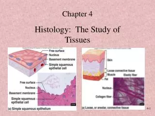
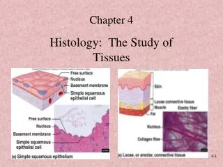
Biology 2011 Practice Histology Lab Practical: PowerPoint Examples.

E N D
Biology 2011Practice Histology Lab Practical:PowerPoint Examples
Each of the following slides has a sample question concerning the microscopic image of a tissue. Select your answer from the choices on the slide. Make your own answer sheet. There are 25 multiple choice questions.An answer key to this practice histology lab practical can be found at the end of this presentation.
#1 Identify the epithelium indicated by the letter A: • Simple squamous • Stratified squamous • Simple cuboidal • Simple columnar • Transitional
#2Identify the cell organelle indicated by the letter B: • Brush border • Cell membrane • Nucleus • Cytoplasm • Vacuole
#3Identify the structure indicated by the letter B: Interstitial lamellae Concentric lamellae Lacuna Central canal Osteon
#4Identify the cell indicated by the letter B: • Leukocyte • Erythrocyte • Phagocyte • Chondrocyte • Thrombocyte
#5Identify the structures indicated by the letter B • Central canal • Concentric lamellae • Canaliculi • Interstitial lamellae • Lacunae
#6 Identify the epithelial tissue type indicated by the letter B a. Simple squamous b. Stratified squamous c. Simple cuboidal d. Simple columnar e. Transitional
#7Identify the epithelial tissue type indicated by the letter B a. Simple squamous b. Stratified squamous c. Simple cuboidal d. Simple columnar e. Transitional
#8Identify the cell organelle indicated by the letter B a. Brush border b. Cell membrane c. Nucleus d. Cytoplsm e. Vacuole
#9 Identify the epithelial layer (stratum) indicated by the bracket labeled A • Corneum • Lucidum • Granulosum • Spinosum • Basale
#10Identify the cell indicated by the letter B: • Leukocyte • Erythrocyte • Phagocyte • Chondrocyte • Thrombocyte
#11Identify the epithelial tissue type indicated by the letter B a. Simple squamous b. Stratified squamous c. Simple cuboidal d. Simple columnar e. Transitional
#12 Identify the structure indicated by the letter B: • Interstitial lamella • Concentric lamella • Lacuna • Central canal • Periosteum
#13 Identify the cells indicated by the letter B • Leukocyte • Erythrocyte • Phagocyte • Chondrocyte • Thrombocyte
#14 Identify the structure indicated by the letter A • Nerve fiber • Hair follicle • Sweat gland • Sebaceous gland • Dermal papilla
#15 Identify the epithelial layer (stratum) indicated by the letter A • a. Corneum • Lucidum • Granulosum • Spinosum • Basale
#16 Identify the tissue type depicted in this slide • Dense fibrous connective tissue • Smooth muscle • Skeletal muscle • Elastic cartilage • Cardiac muscle
#17 Identify the cell structure indicated by the letter B a. Intercalated disc b. Cross striations c. Nucleus d. Cytoplasm e. Vacuole
#18 Identify the tissue type indicated by the letter A • Hyaline cartilage • Epidermis • Compact bone • Adipose tissue • Smooth muscle
#19 Identify the cells indicated by the letter B • Leukocyte • Thrombocyte • Neuron • Chondrocyte • Glial cell
#20 Identify the epithelial layer (stratum) indicated by the letter A • Corneum • Lucidum • Granulosum • Spinosum • Basale
#21 Identify the tissue type depicted in this slide • Dense fibrous connective tissue • Smooth muscle • Skeletal muscle • Elastic cartilage • Cardiac muscle
#22 Identify the cells indicated by the letter B • Skeletal muscle • Cardiac muscle • Smooth muscle • Adipocyte • Keratinocyte
#23 Identify the cell indicated by the letter B • Cardiac muscle • Fibrocyte • Neuron • Chondrocyte • Glial cell
#24 Identify the tissue type indicated by the letter A • Fibrocartilage • Epidermis • Dermis • Adipose tissue • Smooth muscle
#25 Identify the tissue type indicated by the letter B • Nervous • Areolar connective • Dermis • Adipose tissue • Spongy bone
Biology 2011 The End of the Practice Lab Practical:The Answer Key Follows
Key To Practice Histology Practical • C -Simple cuboidal • C - Nucleus • D - Central canal • B - Erythrocyte • E - Lacunae • C - Simple cuboidal • A - Simple squamous • E - Vacuole • A - Corneum • A - Leukocyte • D - Simple columnar • B - Concentric lamella • E - Thrombocyte • C -Sweat gland • B - Lucidum • B - Smooth muscle • A - Intercalated disc • B - Epidermis • E - Glial cell • E - Basale • E - Cardiac muscle • A - Skeletal muscle • C - Neuron • C - Dermis • D - Adipose tissue
Note to 2011 Students Your actual midterm practical will contain PowerPoint slides on more than just histology. There will be charts and diagrams. See your midterm practical review outline for all the topics which will be represented on the midterm practical.