Hyaline change
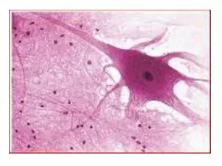
Hyaline change. It is a change in the physical appearance of the tissues which become homogenous, glossy, translucent and pink with H&E staining There is fusion of cells and tissues probably due to coagulation and dehydration of proteins. Types. Three types of hyaline change are recognized

Hyaline change
E N D
Presentation Transcript
Hyaline change It is a change in the physical appearance of the tissues which become homogenous, glossy, translucent and pink with H&E staining There is fusion of cells and tissues probably due to coagulation and dehydration of proteins
Types • Three types of hyaline change are recognized • Keratohyaline • Cellular hyaline • Connective hyaline
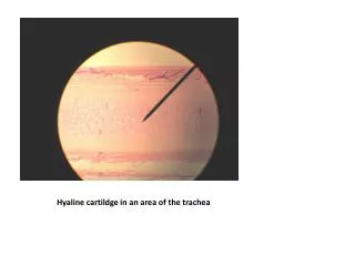
1-Keratohyaline • Excessive keratinization (cornification) of skin occurs in different conditions • Mechanical irritation causes corns and calluses on hands (workmen) , feet (shoes) and place of saddle and harness (horses) • Chlorinated naphthalene poisoning- hyperkeratosis in cattle • Pappiloma virus causes warts in cattle and dogs • Squamous cell carcinoma produces keratohyaline in the form of epithelial pearls • Vit.A deficiency causes keratinization of esophagus and trachea in chicken
2-Cellular hyaline • Cells in many organs are desquamated, particularly in the lumens and cavities of the glands where they fuse into homogenous round masses. • These masses stain deep brown with iodine and are called corpora amylacea. • Commonly found in mammary glands, prostate, lungs, kidneys, and ventricles of brain.
3-Connective hyaline • This is observed in old scars, renal glomeruli in chronic nephritis and in the media of blood vessels in arteriosclerosis.
Significance • Hyaline change occurs under a wide variety of pathological conditions but is not considered significant. It makes the tissues inelastic and lacks nerve and blood supply
General Pathology (PATH 303) Lecture # 5 • Amyloid and Amyloidosis
1. Amyloid and Amyloidosis • Hyaline, waxy material. • Homogenous nature -15 different types. • Physical nature-- non-branching fibrils. Beta-pleated sheet structure (A regular element of secondary structure in proteins, in which two or more extended strands of the polypeptide chain lie side by side (running either parallel or antiparallel), held together by a regular array of hydrogen bonds between backbone NH and C=O groups, to form a ridged planar surface.)
2. Chemical nature of amyloid • 95% fibriller glycoprotein • 5% is non-fibriller glycoprotein ( P component) and proteoglycans • AA ( amyloid associated) – non- immunoglobulin protein synthesized in liver and present in serum- SAA. • Most common form found in animals. • Occurs in chronic inflammatory conditions like T.B. and osteomyelitis- Secondary amyloidosis. • AL (Amyloid light chain) – contains immunoglobulin light chains- Primary amyloidosis. • Produced by plasma cells in plasmacytomas. • Most common form in humans.
3. Occurrence • Most common in man but all species can be affected. • Among animals, common in dog, cattle, horse and chicken. • May be local or generalized.
4. Causes • Secondary amyloidosis in animals is associated with chronic inflammatory disorders. • Activation of macrophages produces cytokines IL-1 and IL-6 • Under the influence of cytokines liver cells produce more SAA. • More SAA alone does not cause amyloidosis • There is enzyme defect in affected animals producing incomplete breakdown of SAA. • Production of insoluble AA protein
Stimulus Unknown (carcinogen ?) Monoclonal B-Lymphocyte proliferation Plasma Cells Immunoglobulin light chains Soluble Precursor Limited Proteolysis Insoluble Fibrils Insoluble AL Protein
5. Primary amyloidosis in man • Deposition of amyloid light chain (AL) or their fragments or both. • Monoclonal cell proliferation- multiple myeloma • Plasma cells also secrete Lamda and Kappa light chains- Bence Jones proteins. • Only a few patients with Bence Jones proteins develop amyloidosis. • Defective proteolysis produces insoluble AL
Chronic Inflammation Stimulus Macrophage activation Interleukins 1 and 6 Liver cells SAA Proteins Soluble Precursor Limited Proteolysis Insoluble Fibrils Insoluble AA Protein
6. Harmful effects: • Pressure atrophy of surrounding cells and tissues • Inhibition of exchange of gases, nutrients and waste materials. • Compression and stenosis of hollow organs.
Gross and microscopic appearance • Amyloid is most commonly seen in spleen, kidney, liver and adrenal. Spleen is normally involved in the chicken with tuberculosis. • In spleen, amyloid gradually forms a cuff around the central artery of spleenic follicles • The mass appears as grey foci, resembling grains of sago- called sago spleen. • The foci gradually increase in size and may rupture and cause fatal hemorrhages.
Special stains • Amyloid stains pink with eosin, yellow with van Gieson, red with Congo red. Crystal violet and methyl violet also stain it red.
Significance • Amyloid deposition is a permanent change and persists for life. • It may cause uremia (in kidney), rupture (in liver) and diabetes in case of pancreas.