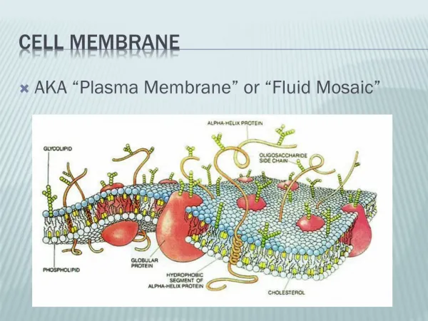
Hyaline Membrane Disease
400 likes | 949 Vues
Hyaline Membrane Disease. Vincent Patrick Uy. Infant’s First Breath. Intermittent compression of the thorax facilitates removal of lung fluids Surfactant DECREASES surface tension to allow low pressure to aerate the lungs – preventing alveolar collapse

Hyaline Membrane Disease
E N D
Presentation Transcript
Hyaline Membrane Disease Vincent Patrick Uy
Infant’s First Breath • Intermittent compression of the thorax facilitates removal of lung fluids • Surfactant DECREASES surface tension to allow low pressure to aerate the lungs – preventing alveolar collapse • Functional residual capacity (FRC) must be established • Air entry into the alveoli displaces fluid, decreases the hydrostatic pressure and increase pulmonary blood flow.
Infant’s First Birth • Decline in PaO2 • Decline in pH • Rise in PaCO2 • Redistribution of the cardiac output • Decrease body temperature • Tactile and sensory inputs
Timeline of Lung Development • Embryonic Period • Protrusion from the foregut • Initial branching • Saccular Stage • Gas exchange may be possible • Septal growth into saccules and into alveoli • Pseudoglandular • 15-20 generations of air branching • Progressive epithelial differentiation • Canalicular • Bronchioles and ducts of gas exchange regions are formed • Alveolar Type II cells 16-33 days AOG 7-16th weeks AOG 16th-25th week AOG >24 weeks AOG
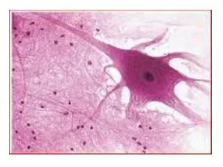
Lung Surfactant • Type II pneumocytes • Reduces surface tension allowing lesser pressures to maintain the alveoli open.
Lung Surfactant • 20 weeks – start to appear (appearance of lamellar bodies) • 28-32 weeks – detectable in amniotic fluid • 35 weeks – “Mature” levels of surfactant
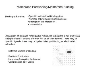
Components of Lung Surfactant • Lipids (70%) • Majority of the lipid component is dipalmitoylphosphatidyl choline (DPPC) which is the major surface tension reducing substance • Proteins (30%) • Hydrophobic surfactant proteins (SP) B & C • Hydrophilic SP A & D
Surfactant Proteins Hydrophobic Hydrophilic
Synthesis, Secretion and Adsorption of Surfactant Tubular Myelin Lamellar body Type II pneumocyte
Factors that Enhance Surfactant Synthesis • Normal pH • Normal temperature of the neonate • Normal perfusion • Adequate amount of oxygen • Low insulin levels • Chronic intrauterine stress (Pregnancy-induced hypertension) • Twin gestations • Antenatal corticosteroids
Hyaline Membrane Disease • Occurs primarily in premature babies; inversely related to gestational age • 60-80% of infants <28 weeks • 15-30% of infants 32-36 weeks • Rare in term neonates (consider genetic abnormalities in surfactant proteins) • Incidence increases with: • Maternal DM • Multiple gestations • Asphyxia • Cold stress • Maternal history of previously affected infants
Pathophysiology • Poor Surfactant Quantity and Quality • Lungs of premature babies have surfactant rich in phosphatidylinositol and smaller amounts of phosphatidylglycerol (PPG). PPG has the greatest surface activity. • Protein content of surfactant from preterm lung is low relative to the amount of phospholipids. • Inflammation and pulmonary edema ensues
Pulmonary Edema • Leads to poor gas exchange • Results from inflammation and lung injury • Reduced pulmonary fluid reabsorption • Low urine output • Proteinaceous edema and inflammatory cytokines increase the conversion rate of surfactant into inactive forms.
Lung Mechanics in Preterms • Worsening RDS with formation of hyaline membranes result in less compliant lungs • Lower part of the chest is pulled in as the diaphragm descends intrathoracic pressure is more negative atelectasis • Highly compliant chest wall less resistant volume of the lung tends to approach RV Atelectasis
Disease Processes Low surfactant levels ATELECTASIS HYPOXEMIA Chest Compliance Small Alveoli HYPERCAPNIA Alveolar Ventilation Impaired Pulmonary Artery Constriction Shunting Ischemic Injury to the lungs Pulmonary Artery Constriction Proteinaceous effusion into the alveolar space
Clinical Manifestations HISTORY PHYSICAL EXAM Tachypnea Grunting Nasal flaring Retractions Cyanosis Decreased breath sounds • Often preterm • Had asphyxia in the perinatal period • Respiratory distress at birth • Apnea **Classic chest radiograph is also an additional feature of the disease.
Diagnostic Tests • Chest Radiograph • Blood Gas sampling • Sepsis Work-up • Serum glucose levels • Serum electrolytes and calcium levels • Echocardiography
Preventive Management • Avoid unecessary and untimely Cesarean sections. • Antenatal Corticosteroids – 24-34 weeks gestation – is associated with overall reduction in neonatal deaths, RDS, IVH, NEC, ICU admissions and systemic infections in the first 48 hours of life • Betamethasone: Two 12 mg doses IM given 24 hours apart • Dexamethasone is no longer given due to increase risk of cystic periventricular leukomalacia among preemies
Surfactant Replacement • Considered the standard of care in RDS • Surfactant prophylaxis (within 15 minutes of birth) to all infants <27 weeks. • Consider prophylaxis if 27-29 weeks if baby was intubated or mother did not get antenatal steroids • Repeated doses every 6-12 hours for a total of 3-4 doses.
Natural Surfactant • Obtained from animal lung lavage or by mincing lung tissues • Lipid extraction removes hydrophilic components (SP-A and SP-D). The purified lipid derivative contains the necessary components to control the surface tension • Choice of natural surfactant is based on clinician/hospital preference
Respiratory Management • Because of increase risk of BPD, preterm infants without signs of respiratory failure can be managed with CPAP or NIPPV
Respiratory Management • Indications for immediate intubation and mechanical ventilation: • Respiratory acidosis (pH <7.20 and PCO2 >60 mmHg) • Hypoxemia • Severe apnea • Unresponsive and limp babies with impending respiratory distress
Target Values • Oxygen Saturation • Saturations above 95% and below 89% are associated with poor outcomes • O2sat by Pulse ox – 90-95% is optimal • PCO2 levels • 45-60 mmHg is the optimal level • If it exceeds 60 mmHg, the pH falls <7.25 which is associated with poor CV function • Babies initially on CPAP that develop acidosis, should be intubated
Sedation and Pain Relief Advantages Disadvantages Side effects Morphine – hypotension Fentanyl – rigid chest wall Benzo’s – Tolerance and dependence • Improved ventilatory synchrony and pulmonary function • Neuroendocrine responses are alleviated • Decreased adverse long term neurologic sequelae
Supportive Measures • Umbilical artery line • Thermoregulation • Fluid management • Treat hypotension with vasopressor support and cautious use of saline boluses • Early nutrition
Complications • Survival from HMD is dependent of gestational age and birthweight • Major morbidities such as IVH, BPD and NEC remain high in smaller infants • Endotracheal tube complications • Air leak syndromes – rupture of overdistended alveoli • BPD
Bronchopulmonary Dysplasia • Result of lung injury among infants managed with mechanical ventilation and supplemental oxygen • Defined as persistent oxygen dependency up to the 28 day of life.