Back Pain
Back Pain. Tanya Potter Consultant Rheumatologist. Case of Back Pain. 34 year old lady on post-natal ward Admitted with left sided lumbosacral pain Relevant questions/thoughts?. Case of Back Pain. 4 th pregnancy, uncomplicated delivery, no epidural


Back Pain
E N D
Presentation Transcript
Back Pain Tanya Potter Consultant Rheumatologist
Case of Back Pain • 34 year old lady on post-natal ward • Admitted with left sided lumbosacral pain • Relevant questions/thoughts?
Case of Back Pain • 4th pregnancy, uncomplicated delivery, no epidural • Night and rest pain, left thigh radiation, worse with movement, unable to walk or weight bear • Episode of feeling cold and shivery 4 days previously
Case of Back Pain • OE in pain, not unwell, afebrile, haemodynamically stable • Tender left lumbosacral region, unable to do SLR • Hip ok • No neurology, bladder and normal PR
Case of Back Pain • ESR 101, CRP 201 • ALP 489, ALB 19 • Hb 9.8 MCV 76 • Differential diagnosis and further investigations?
MRI septic arthritis of left SIJ with an abscess in thegreater sciatic notch
Blood cultures Beta-haemolytic Strep. • IV Antibiotics • Orthopaedic review • CT guided aspiration • Few weeks later CRP 28 • Repeat MRI
Some resolution of abscess, marrow oedema, some destruction of SIJ MRI
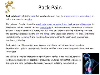
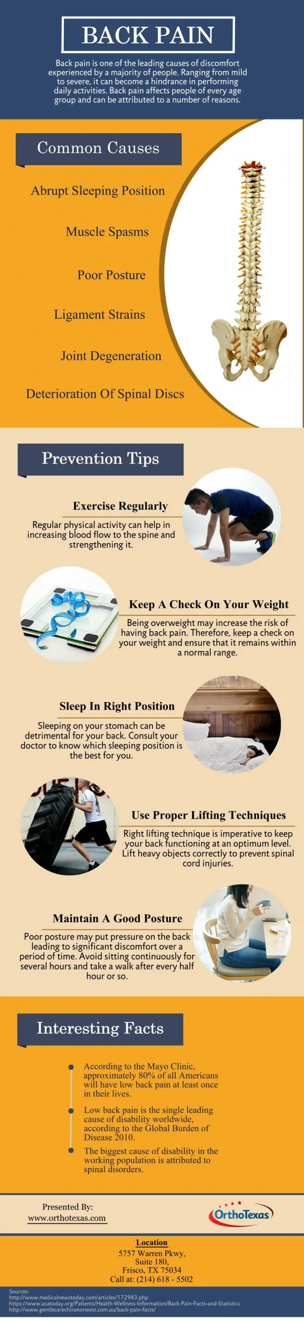
Causes • Simple mechanical eg ligamentous strain • Degenerative disease with/without neural, cord or canal compromise • Metabolic – osteoporosis, Pagets • Inflammatory – Ankylosing spondylitis • Infective – bacterial and TB • Neoplastic • Others, (trauma, congenital) • Visceral
Triggers to investigate/ refer • Red Flags ?
Red flags • Age <20 or >50 with back pain for the 1st time • Thoracic pain >50 yrs • Pain following a violent injury/trauma • Unremitting, progressive pain
Red flags • Past or current history of cancer • On Steroids or immunosuppressants • Drug abuser or +ve HIV • Systemic symptoms - fever, appetite and weight loss, malaise
Red flags • Bilateral leg radiation, sensory/motor/sphincter symptoms • Pain predominantly at night
Inflammatory flags • Morning stiffness and pain >30 mins -1 hr • Better with activity • Peripheral joint involvement • Anterior uveitis • Psoriasis • Inflammatory bowel disease • Recent GI or GU infection • Family history
Myotomes • C5 Deltoid, biceps (biceps jerk) • C6 Wrist extensors, biceps (biceps, brachioradialis jerk) • C7 Wrist flexors, finger extensors, triceps (triceps jerk) • C8 Finger flexor, thumb extensors (triceps jerk) • T1 finger abductors
Myotomes • L2 Hip flexion • L3 Knee extension (knee jerk) • L4 Knee extension, ankle dorsiflexion (knee jerk) • L5 toe dorsiflexion • S1 foot plantar flexion, eversion
Examination • LOOK – deformity, muscle wasting, kyphosis, scoliosis • LOOK – normal cervical lordosis, thoracic kyphosis, lumbar lordosis • FEEL – spinal processes and sacroiliac joints
Examination • MOVE – Lumbar flexion • Schober’s test – marks at “dimples of Venus” and 10 cm above. Measure at maximal flexion – usually 5 cm • MOVE – Lumbar lateral flexion • MOVE – Cervical flexion/extension, lateral rotation and flexion, thoracic rotation
Examination • Sciatic stretch (patient supine) - Straight leg raise and dorsiflexion of foot - pain in calf and posterior thigh between 30-70o – low lumbar (L5/S1) lesion or sciatic irritation • Femoral stretch (patient prone) – knee is flexed and then hip extended – pain in anterior thigh – high lumbar (L2-L4) lesion
Imaging • XR – • Isotope Bone scan – • MRI – • CT
Imaging • XR – tumour, fracture, infection, inflammation • Isotope Bone scan – • MRI – • CT
Imaging • XR – tumour, fracture, infection, inflammation • Bone scan – increased turnover eg infection, metastatic disease, fractures, Pagets • MRI – • CT
Imaging • XR – tumour, fracture, infection, inflammation • Bone scan – increased turnover eg infection, metastatic disease, fractures, Pagets • MRI – soft tissue, discs, facet joint, nerve roots, cord, inflammation • CT
Degenerative disease and sciatica • Very common • Facet joint OA, disc disease, osteophyte • Mechanical back pain • Sciatica – most resolve NB persistent, neurology, bilateral, red flags • Analgesia, PT, pain clinics
Malignancy • Which cancers associated with bone mets?
Malignancy • Unremittting, progressive and night pain • Systemic symtoms • Past hx Ca • Breast, bronchus, thyroid, kidney, prostate and myeloma/plasmacytoma • Osteolytic (prostate osteoblastic) • XR can be normal in early stages – further imaging if suspicion high • Predilection for vertebral body and pedicles
Infection • discitis, osteomyelitis, and epidural abscess. • hematogenously spread • most often Staphylococcus aureus. • Gram-negative rods in postoperative or immunocompromised patients • normal skin flora is less commonly isolated in postoperative patients. • Postoperative patients develop symptoms 2 to 4 weeks after surgery after an initial improvement in pain.
Infection • Pseudomonas organisms • Mycobacterium tuberculosis in developing nations and immigrant population. Fungal infections are rare. • Only one third have fever and 3% to 15% present with neurologic deficit. • Infections typically involve the intervertebral disc and vertebral body endplate
Infection • Radiographic changes at 2 to 4 weeks • bone scan can be positive as early as 2 days 75% specific • MRI appearance is decreased T1- and increased T2-weighted signal in the infected disk. Enhancement after gadolinium
Infection • Conservative treatment of antibiotics, rigid bracing to prevent deformity and control pain • Surgery : neurologic deficit, presence of abscess, extensive bone loss with kyphosis and instability, failure of blood work and biopsy to isolate any organism, excision of a sinus tract, or no response to conservative treatment.